ZOO 3000 Study Guide - Final Guide: Corneal Endothelium, Loose Connective Tissue, Vitreous Body

Document Summary
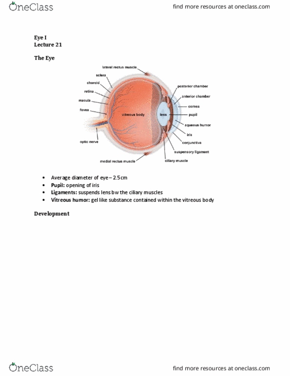
The wall of the eye: the wall of the eyeball is organized into three layers of tissue, cornealscleral coat, cornea and sclera. Structural role: vascular coat, choroid, ciliary body, and iris. Can"t have bv where light enters the eye: retina, retinal pigment epithelium (rpe) and neural retina tissue with photoreceptors. Rpe covers retina between retina and vascular coat. Does not impede the passage of light. Replaced and shed: bowman"s membrane (basal lamina, stroma (dense regular connective tissue) Important for focusing light on the fovea: descemet"s membrane (basal lamina, corneal endothelium. Provides for metabolic exchange between cornea and aqueous humour as the cornea is not vascularized: tear fluid keeps the cornea from dehydrating and helps give nutrients. Choroid layers: choriocapillary layer (ch, loose connective tissue and fenestrated capillaries, supplies nutrients to retina, lamina vitrea/bruch"s membrane (lv, thin layer between capillaries and pigment epithelium of the retina, separates vascular coat from retina.