BIOL 204 Study Guide - Midterm Guide: Yolk Sac, Somite, Blastula


Definition/Function
Zygote - single fertilized egg
-
Cleavage - rapid division
-
Morula - almost solid ball
-
Blastula Formation - Hollow ball of cells
-
Blastocoel - hollow space inside the blastula
-
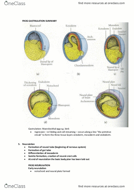
Gastrulation
Differentiation of ectoderm, mesoderm, and endoderm
○
Cells from outer surface migrate inside the embryo via Blastopore
(opening)
○
Formation of the archenteron
Becomes the lumen of the digestive tract
§
○
Endoderm will eventually become the lining of the digestive tract
○
-
Neurulation
Forms Neural Tube and Neural Crest
○
Somite formation in the trunk region (epimere mesoderm)
Arranged Anterior to Posterior
§
○
-
Somite
Blocks of epimere mesoderm
○
Visible underneath the dorsal layer of ectoderm
○
-
Microlecithal - Little yolk
-
Mesolecithal - Medium amount of yolk
-
Macrolecithal - Lots of yolk
-
Mesenchyme
special type/ undifferentiated mesodermal cells that are not linked
together into sheets and can move independently --> migrating
rapidly
○
Anterior is more developed than posterior
○
-
Animal Pole - Lower concentration of yolk
-
Vegetal Pole - higher concentration of yolk
-
Vitelline Vessels - transport blood and receive nutrients from yolk sac
-
Anatomical distinctions
Dorsal - Back
-
Ventral - belly
-
Medial - Close to midline
-
Lateral - Away from midline
-
Anterior - Closer to head
-
Posterior - Closer to tail
-
Proximal - Towards attachment site
-
Distal - Away from attachment site
-
Embryology Origins
Mesoderm differentiates into:
Epimere mesoderm
In trunk region
§
Made of somites - discrete blocks
Dermatome □
Myotome□
Sclerotome □
§
-
Mesomere mesoderm
-
Hypomere mesoderm
Splanchnic hypomere - inner layer
§
Somatic hypomere - outer layer
§
-
-
Made of Mesoderm
Notochord
-
-
What occurs at different life stages of chick embryo?
33 hr chick
pair of dorsal aortas
§
Circulatory system starting to form
§
-
48 hr chick
Distinct blood vessels
§
Vitelline vessels
§
Extra embryonic membranes forming
Somatopleure (ectoderm and somatic hypomere
mesoderm)
Amnion
®
Chrion
®
□
Splanchnopleure (endoderm and splanchnic hypomere
mesoderm)
Yolk sac
®
Allantois
®
□
§
-
-
Morphological Differences
Yolk Distribution
Amphioxus egg --> microlecithal
-
Frog egg --> mesolecithal
-
Chick egg --> macrolecithal
-
-
Archenteron Formation
Entercoely
Mesoderm from dorsal lining of archenteron forms 2 pouches
§
Space in the middle forms a coelom and the lining is made
from mesoderm
§
Coelom forms main body cavity (major organs in the adult)
§
-
-
Location and How to Identify
Neural Plate - mid dorsal region of ectoderm
-
Notochord - ventral to the neural plate
-
Figures to Draw:
Fig 1. Developmental process
Fig 2. Sagittal Section of amphioxus during Gastrulation
Fig 3. Neurulation of the Amphioxus
Fig 4. Close up of Neurulation
Fig 5. Cross Section of Frog embryo
Fig 6. External chick embryo
Fig 7. 4 Cross sections of Chick embryo
Fig 8. Cross section from 33 hour chick embryo
Fig 9. Full mount of 48 hr chick embryo
Lab 1 - Embryology
February 11, 2018
3:00 AM

Definition/Function
Zygote - single fertilized egg
-
Cleavage - rapid division
-
Morula - almost solid ball
-
Blastula Formation - Hollow ball of cells
-
Blastocoel - hollow space inside the blastula
-
Gastrulation
Differentiation of ectoderm, mesoderm, and endoderm
○
Cells from outer surface migrate inside the embryo via Blastopore
(opening)
○
Formation of the archenteron
Becomes the lumen of the digestive tract
§
○
Endoderm will eventually become the lining of the digestive tract
○
-
Neurulation
Forms Neural Tube and Neural Crest
○
Somite formation in the trunk region (epimere mesoderm)
Arranged Anterior to Posterior
§
○
-
Somite
Blocks of epimere mesoderm
○
Visible underneath the dorsal layer of ectoderm
○
-
Microlecithal - Little yolk
-
Mesolecithal - Medium amount of yolk
-
Macrolecithal - Lots of yolk
-
Mesenchyme
special type/ undifferentiated mesodermal cells that are not linked
together into sheets and can move independently --> migrating
rapidly
○
Anterior is more developed than posterior
○
-
Animal Pole - Lower concentration of yolk
-
Vegetal Pole - higher concentration of yolk
-
Vitelline Vessels - transport blood and receive nutrients from yolk sac
-
Anatomical distinctions
Dorsal - Back
-
Ventral - belly
-
Medial - Close to midline
-
Lateral - Away from midline
-
Anterior - Closer to head
-
Posterior - Closer to tail
-
Proximal - Towards attachment site
-
Distal - Away from attachment site
-
Embryology Origins
Mesoderm differentiates into:
Epimere mesoderm
In trunk region
§
Made of somites - discrete blocks
Dermatome
□
Myotome
□
Sclerotome
□
§
-
Mesomere mesoderm
-
Hypomere mesoderm
Splanchnic hypomere - inner layer
§
Somatic hypomere - outer layer
§
-
-
Made of Mesoderm
Notochord
-
-
What occurs at different life stages of chick embryo?
33 hr chick
pair of dorsal aortas
§
Circulatory system starting to form
§
-
48 hr chick
Distinct blood vessels
§
Vitelline vessels
§
Extra embryonic membranes forming
Somatopleure (ectoderm and somatic hypomere
mesoderm)
Amnion
®
Chrion
®
□
Splanchnopleure (endoderm and splanchnic hypomere
mesoderm)
Yolk sac
®
Allantois
®
□
§
-
-
Morphological Differences
Yolk Distribution
Amphioxus egg --> microlecithal
-
Frog egg --> mesolecithal
-
Chick egg --> macrolecithal
-
-
Archenteron Formation
Entercoely
Mesoderm from dorsal lining of archenteron forms 2 pouches
§
Space in the middle forms a coelom and the lining is made
from mesoderm
§
Coelom forms main body cavity (major organs in the adult)
§
-
-
Location and How to Identify
Neural Plate - mid dorsal region of ectoderm
-
Notochord - ventral to the neural plate
-
Figures to Draw:
Fig 1. Developmental process
Fig 2. Sagittal Section of amphioxus during Gastrulation
Fig 3. Neurulation of the Amphioxus
Fig 4. Close up of Neurulation
Fig 5. Cross Section of Frog embryo
Fig 6. External chick embryo
Fig 7. 4 Cross sections of Chick embryo
Fig 8. Cross section from 33 hour chick embryo
Fig 9. Full mount of 48 hr chick embryo
Lab 1 - Embryology
February 11, 2018 3:00 AM