ANP 1107 Study Guide - Midterm Guide: Descending Limb Of Loop Of Henle, Passive Transport, Transport Protein

11 Mar 2017
School
Department
Course
Professor
Document Summary
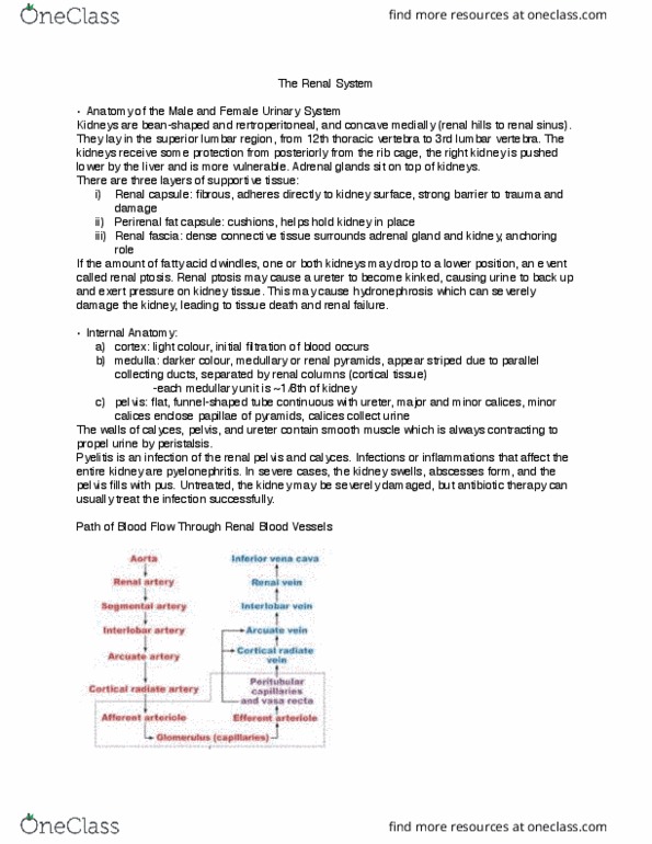
Superior lumbar region from t12 to l3. Right kidney pushed lower by the liver. Adrenal glands sit on top of the kidneys. Right kidney is surrounded by an extra layer of adipose tissue of brown fat, covered by peritoneum a retroperitoneal organ. Renal capsule: fibrous, adheres directly to kidney surface and attaches it and the adrenal gland to surrounding structures. Perirenal fat capsule: cushions, helps hold kidney in place. Renal fascia: dense ct surrounds adrenal gland & kidney anchoring role. Renal ptosis: event when kidneys drop to a lower position when the fatty tissue dwindles (perirenal fat capsule) Contain medullary/ renal pyramids separated by renal columns (cortical tissues: the medullary are stripped because they are formed by parallel bundles of microscopic urine collecting tubules. Pyelitis: infection of the renal pelvis and calyces. Pyelonephritis: when the bacteria and infection affects the entire kidney. Renal arteries: deliver one-fourth of the total cardiac output to the kidneys.