PSYB64H3 Study Guide - Midterm Guide: Positron Emission Tomography, Transcranial Magnetic Stimulation, Pia Mater

10 Jun 2016
School
Department
Course
Professor
Document Summary
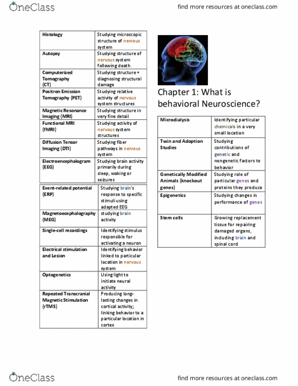
Histology: study of cells and tissues at the microscopic level. Autopsy: the examination of body tissues following death. Computerized tomography (ct): computers are used to study structure and diagnose structural damage through x-rays of the brain. Positron emission tomography (pet): observe brain activity through radioactive atoms. Magnetic resonance imaging (mri): studying the brain through very high resolution structural images. Functional mri (fmri): assess activity of the nervous system. Diffusion tensor imaging (dti): using mri technology to trace fiber pathways in the brain. Electroencephalogram (eeg): studying brain activity through electrodes placed on the scalp, usually during sleep and waking or seizures. Event-related potentials (erps): studying the brain"s response to specific stimuli using an adapted eeg. Magnetoencephalography (meg): recording of the brain"s magnetic output activity. Single-cell recordings: recording the activity of single neurons through surgically implanted microelectrodes. Electrical stimulation: identifying behavior linked to a specific area in the nervous system and brain.