MGY277H1 Study Guide - Midterm Guide: Lipid Bilayer, Catabolite Activator Protein, Repressor

8 Jun 2018
School
Department
Course
Professor

MGY277 UNIT 2
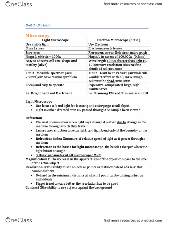
• Microscopy Principles
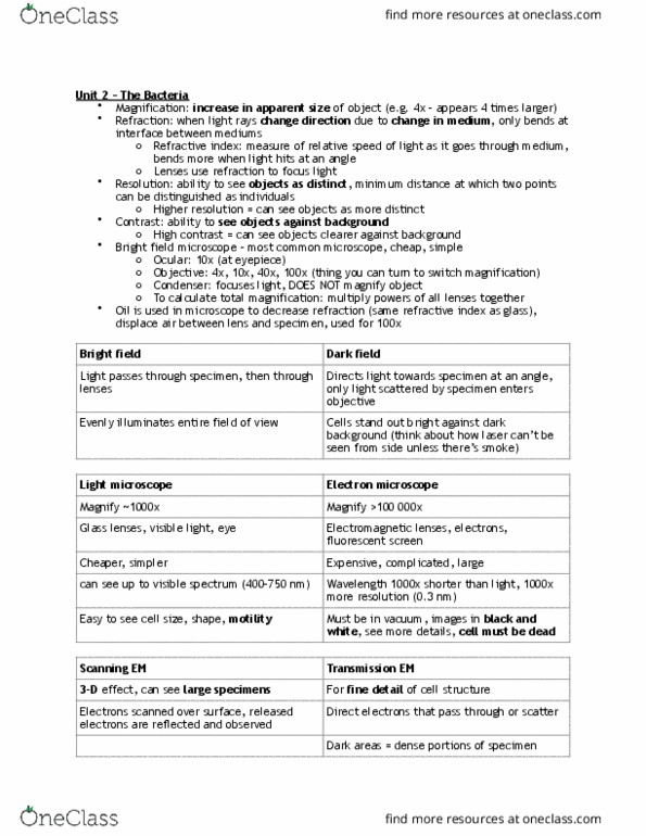
- Magnification: The increase in the apparent size of the object compared to the size of
the actual object.
o i.e. how much larger you made the object with the lenses of the microsope.
- Resolution: The ability to observe the objects (or points) as distinct and separate (good
resolution), instead of as one large blur (poor resolution).
- Contrast: The ability to see objects against the background.
o i.e. you will be able to see the objects clearly with high contrast
o Some techniques to increase contrast include: dark field microscopy and staining.
- Refraction: A phenomenon in which light rays change direction due to a change in the
medium through which they travel (e.g. water, air, oil).
o Light microscope utilizes lenses to refract light in order to focus and magnify the
objects.
o Refractive index: measure of relative speed of light as it passes through a
medium.
▪ i.e. Refractive indexes for air and water are different, which is why the
pencil looks broken in a glass of water (PWN’s example).
- Light Microscope
o Bright-field microscope
▪ Most common
▪ Light travels through the specimen and then lenses
▪ Two types of lenses: objective (directly above the specimen; 4x, 10x, 40x
and 400x) and ocular (the part where we look into; 10x).
• Total magnification = magnification of objective lens * magnification
of ocular lens
▪ Condenser can focus/disperse the light
▪ Resolution: maximum 0.2 microns
▪ Unable to see virus
o Dark-field microscope
▪ A modification of bright field; used to improve contrast
▪ Light at an angle → background light does not directly enter objective lens
- Electron Microscope (compared to light microscpes…)
o Electrons replace light
o Electromagnets replace glass lenses
find more resources at oneclass.com
find more resources at oneclass.com

o Resolution and magnification (100,000x) are higher
o Lenses and specimen must be in vacuum
• Gram Staining
- Sample is first spread on a glass slide
- Fixation (using flame): quickly pass the slide through flame
- Slide is washed with crystal violet making the specimen purple
- Slide is then washed with iodine (a mordant) which fixes crystal violet to the specimen
- Slides are rinsed using alcohol (a decolourizer) to remove excess crystal violet. Gram-
positive cells will remain purple; gram-negative cells become colourless.
- Slides are flooded with safranin (a counterstain). Gram-positive cells remain purple;
gram-negative cells appear pink.
- --
- Gram stain utilizes the fundamental differences in cell wall composition between gram
negative and gram positive bacteria.
o The thick, dehydrated peptidoglycan layer of gram positive bacteria appears to
be a permeability barrier, preventing the loss of crystal violet iodine complex
from the alcohol wash.
o Peptidoglycan in gram negative bacteria is thin and large pores. Alcohol extracts
the lipids and increases porosity of the layer, which makes it easier to remove
crystal violet iodine complex.
• Different types of staining
- Simple stains
o One basic dye is used to stain cells.
- Differential stains: a multi-step, multi-dye procedure to stain cells.
o Gram stain
▪ Used to distinguish between gram-positive and gram-negative bacteria. 2
different dyes are used.
o Acid-fast stain
▪ Used to detect organisms that do not easily take up stains (e.g.
Mycobacterium)
- Special Stains
o Capsule stain
▪ Negative stains; take advantage of the fact that viscous capsules do not
take up stains; dye the background and capsules pop out.
find more resources at oneclass.com
find more resources at oneclass.com
Document Summary
Magnification: the increase in the apparent size of the object compared to the size of the actual object: i. e. how much larger you made the object with the lenses of the microsope. Resolution: the ability to observe the objects (or points) as distinct and separate (good resolution), instead of as one large blur (poor resolution). Contrast: the ability to see objects against the background: i. e. you will be able to see the objects clearly with high contrast, some techniques to increase contrast include: dark field microscopy and staining. Resolution: maximum 0. 2 microns: dark-field microscope, a modification of bright field; used to improve contrast, light at an angle background light does not directly enter objective lens. Electron microscope (compared to light microscpes : electrons replace light, electromagnets replace glass lenses, resolution and magnification (100,000x) are higher, lenses and specimen must be in vacuum, gram staining. Sample is first spread on a glass slide.