PSL300H1 Study Guide - Final Guide: Myosin Head, Sarcomere, Myofibril

Document Summary
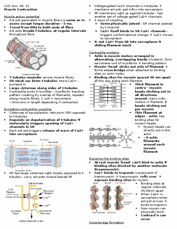
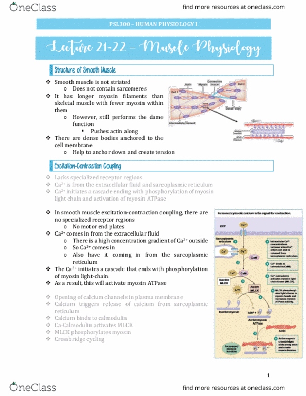
Excitation-contraction coupling : cisternae of the sarcoplasmic reticulum (sr) are apposed to the t-tubules, bringing the ap to the interior of the muscle fibre. Depolarization of the t-tubules, molecularly triggers opening the ca2+ channels in the sarcoplasmic reticulum (voltage dependent). Each ap triggers release of a wave of ca2+ in sarcoplasm. Contractile proteins of the sarcomere: actin (thin filament) and myosin (thick filaments, allows straition appearance of muscle) molecules, arranged in alternating overlapping bands, form the sarcomere unit of myofibrils. Myosin head forms cross-bridge when attached to binding sites on the actin. Myofibrils: muscle cells arranged into myofibrils: single, linear continuous stretch of interconnected sarcomeres, extended length of the muscle cells. Arranged in parallel in the cell to generate more force. ***contractile proteins: sarcolemma: cell membrane, sarcoplasm: muscle cell cytoplasm, sarcoplasmic: stores calcium until it is released for muscle contraction (modified er, t-tubules : allows for spread of muscle excitation deep into muscle, sarcomeres: regular arrangement of myofibrils.