Anatomy and Cell Biology 3309 Study Guide - Midterm Guide: Antigen Presentation, Caveolae, Chondrogenesis

17 Jan 2014
School
Department
Professor


Document Summary
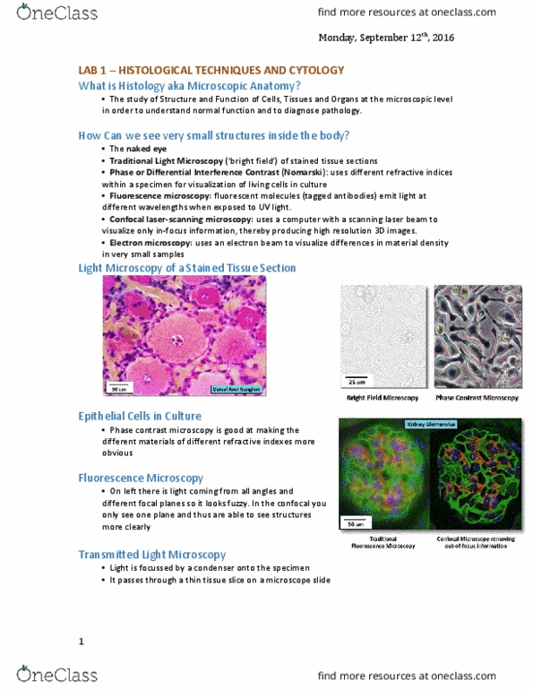
Lecture 1:mammalian histology - introduction histology: microscopic anatomy. Study of tissues at the light and e-microscope level. Ability to visualize structures not normally seen with the naked eye. Samples examined are 2d representations of the original tissue, you must imagine the 3d structure. Tissues undergo chemical modification that can misrepresent the living tissue= artifact. Immunocytochemistry staining method: used formaling bc allows proties to react w/ ab"s. Use ab"s to attach to fluorchromes (fluorescent dyes) which attach to antigens (proteins) Seen with a fluorescent microscope + uv light which activates the dye and emits the color. Dehydrate tissue then infiltrate with liquid embedding soln" to harden and subsequently section. Can be many sections picture of cuts on a tube e. g. figure 8 is at a bend. Section at 60nm (epoxy resin)/ 80nm (plastic) Place on copper grid for e- to pass through. Reveals the cell"s ultrasection (fine structures only visible with an e- microscope)