Biology 1002B Study Guide - Final Guide: Color Vision, Trichromacy, Rhodopsin

19 Apr 2017
School
Department
Course
Professor

Document Summary
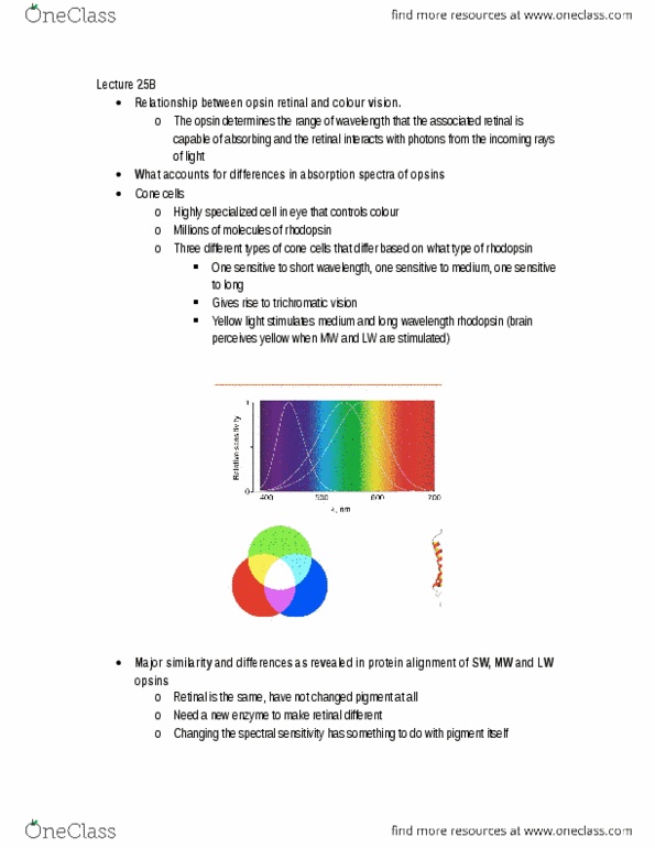
Trichromatic vision: three types of cones in the retina, each with different rhodopsin (pigment) but they all bind to the same retinal: blue (short wavelength), green (medium wavelength), yellow (long wavelength) It is possible to match all colours of the visible spectrum by mixing these 3 colours. Trichromats: organisms with 3 different rhodopsins: can distinguish colour better than organisms with less (dichromats, monochromats) Lysine: specific highly conserved amino acids/residues that bind to retinal. Glutamic acid stabilizes the complex: the protein is much larger than retinal -> stronger influence. Importance of lysine at position 296 and glutamic acid at position 113 of opsins. Lysine at 296: the location of retinal binding. Glutamic acid at 113: stabilizes the lysine retinal bond: hydrogen bonding between n+ and amino acids (-) Association between trichromacy, dichromacy, old world monkeys and new world monkeys. Trichromacy: 3 different opsins: old world (asia, africa, europe) monkeys, and humans, developed trichomacy due to gene duplication.