Health Sciences 2300A/B Study Guide - Final Guide: Coronary Sinus, Circumflex, Pulmonary Vein

29 Jun 2018
School
Department
Course
Professor


The Heart – Exam Study Guide
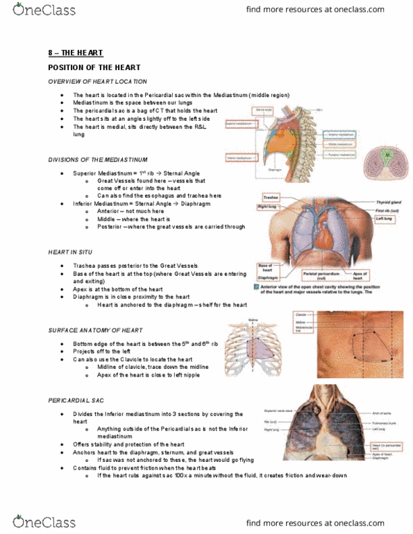
Where is the heart located?
In the pericardial sac within the mediastinum - sits at an angle off to the left side.
Where are the superior, inferior, anterior, middle, and posterior mediastinum located?
Superior: 1st rib - sternal angle
Inferior: Sternal angle to diaphragm
Anterior: Between sternum and pericardium
Middle: Surrounds the pericardium
Posterior: Posterior pericardial wall
What landmarks are used to locate the heart in clinical settings?
- The ribs and clavicle are used – bottom edge of heart sits b/w 5th and 6th rib
- Mid clavicular line aligns with the apex of the heart
What is the purpose of the Pericardial Sac?
- Divides the inferior mediastinum into 3 sections covering the heart
- Anchors heart to diaphragm, sternum, and great vessels
- Contains fluid to prevent friction when heart beats
What are the three pericardial layers?
Fibrous Pericardium
Serous Pericardium
Pericardial Cavity
find more resources at oneclass.com
find more resources at oneclass.com

Clinical Correlate: A baby is born and doesn't seem to be getting enough oxygen or blood flow - what could
have happened?
- The foramen ovale may have failed to close
- This is often caught before birth
- It is meant to remain open while the child is in utero – so you wouldn’t really be able to identify whether it
will close after birth
- Deoxygenated blood mixes w/ oxygenated blood
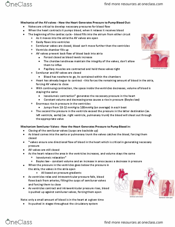
Explain how atrioventricular valves function.
- Function like a parachute
1. AV valve is open and the cusps are open (tricuspid valve) – the chordae tendineae don’t have much
tension and papillary muscles are relaxed
2. Valve is open and blood flows into the ventricle – the increase in pressure forces the valves to close
3. Must prevent backflow from ventricle to atrium
4. Papillary muscles contract putting tension through the chordae tendineae which anchor the valves shut and
prevent them from inverting and letting the blood get back into the atrium
Explain how semilunar valves function.
1. As the pressure builds and increases, the ventricles contract and force the semilunar valves to open.
2. As the blood gets pushed out the first bit of blood is under the highest pressure and will go the farthest (to
toes and finger tips).
3. The last amount of blood isn’t under high pressure and won’t make it over the arch of the aorta or through
the pulmonary arteries.
4. Results in a backflow of blood.
find more resources at oneclass.com
find more resources at oneclass.com
Document Summary
In the pericardial sac within the mediastinum - sits at an angle off to the left side. The ribs and clavicle are used bottom edge of heart sits b/w 5th and 6th rib. Mid clavicular line aligns with the apex of the heart. Divides the inferior mediastinum into 3 sections covering the heart. Anchors heart to diaphragm, sternum, and great vessels. Contains fluid to prevent friction when heart beats. The foramen ovale may have failed to close. It is meant to remain open while the child is in utero so you wouldn"t really be able to identify whether it will close after birth. Semilunar aortic valve cannot close properly due to plaque buildup on the valves. The blood flows back into the ventricle because the valve cannot close properly. Caused by consuming cholesterol that combines w/ substances and forms plaques. Explain the flow of blood in pulmonary circulation. Explain the flow of blood in systemic circulation.