Kinesiology 3337A/B Study Guide - Midterm Guide: Precordium, Epicenter, Cardiac Muscle

10 Dec 2016
School
Department
Course
Professor

Document Summary
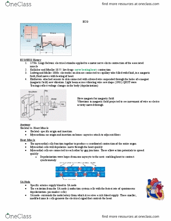
**what you see on paper is the relation between the vector that is being depolarized by the heart and the axis of the ecg lead (which is generated by the unit/machine) Both leads in same direction = biggest depolarization wave (qrst section) As it moves away from parallel (oblique 45 degrees) = the size (smaller depolarization wave) Both leads are perpendicular = no deflection at all (no tracing present) As it moves away from parallel in the opp. direction = smaller amplitude in negative leads parallel in opp. direction = large amplitude in the negative direction direction. Different amplitude waves will be reflected by the different leads therefore this enables you to look through the heart at different angles to see if the electrical activity in that area has been compromised. More tissue that is being depolarized the greater the deflection it will be.