HTH 100 Study Guide - Midterm Guide: Venae Cavae, Beta Blocker, Subarachnoid Hemorrhage


Health Exam 2 Notes
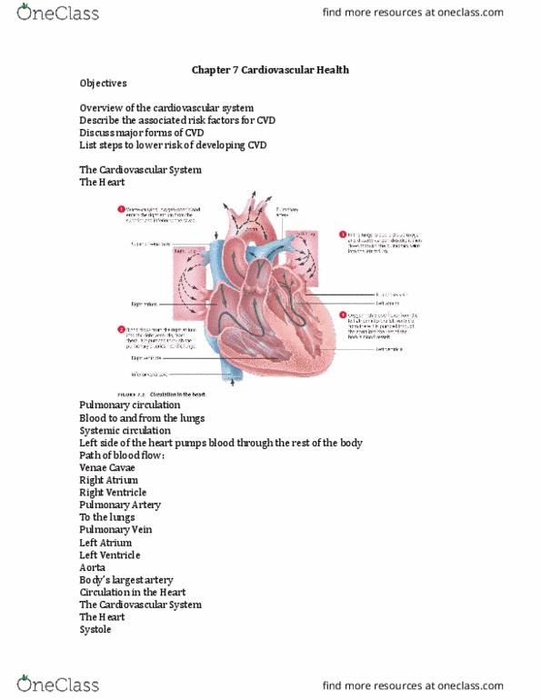
Pulmonary circulation – to lungs and back
Systemic circulation – heart to body and back
Aorta – largest artery in body
Sinus node – pacemaker; receives main signals
Systole – numerator of BP; contracting
Diastole – denominator of BP; relaxing
Capillaries – unite vessels going from veins, arteries, etc.
- From arterioles to venules
Vena cava – largest vein
Arrhythmias – irregularities in heart rhythm
Atrial fibrillation – if blood is not flowing smoothly, it may clot
Ventricular fibrillation – use of paddles; heart cannot pump (deadly)
Cholesterol – fatty, wax-like substance carried in bloodstream
LDL – bad cholesterol; takes cholesterol to organs
HDL – good cholesterol; takes unwanted cholesterol to liver
Pulmonary arteries – carry deoxygenated blood
Plaque – deposits in arteries
Ischemia – reduced oxygen supply to body part/organ
Aneurysm – weakened blood vessel that may bulge/burst
Metabolic syndrome – simultaneous metabolic conditions increasing CVD risks
Carcinogen – cancer-causing agents
Metastasize – cancerous cells spreading to different areas of body
Tumor – neoplasmic mass forming cell clump
find more resources at oneclass.com
find more resources at oneclass.com

Cardiovascular Health
• CVD – Cardiovascular disease
• Heart disease #1 cause of death in U.S. for all age groups
o High rate caused by lifestyles
• Risk Factors:
o Genetics and lifestyle choices
• Consists of heart and vascular system
• Left side of heart pumps to body, right side pumps to lungs
Arteries always go away from the heart (thicker than veins); Veins always carry blood to the
heart
Know the main anatomical structures involved and the path of blood flow through the heart.
1. Deoxygenated blood enters right atrium after circulation through body
2. Blood moves to right ventricle, is pumped through pulmonary artery to lungs, receiving
oxygen
3. Oxygenated blood returns to left atrium
4. Blood from left atrium moves to left ventricle, which pumps blood through aorta to all
body parts
Atherosclerosis:
- Plaque in inner linings of an artery
- 1/3 of all deaths
- Lining damaged by:
o Hypertension, high insulin, and hyperlipidemia, tobacco
- May lead to:
o Stroke, heart attack, and peripheral vascular disease, CAD/CHD
- Blood clots:
o Thrombus
o Embolus (travelling)
o Ischemia (blood flow cut off from tissue)
- Blockage of coronary artery heart attack
- Inner lining damaged by:
o Hypertension
o Hyperlipidemia
o Hyperglycemia
o High insulin levels
find more resources at oneclass.com
find more resources at oneclass.com

Hypertension:
- “ilet Killer – ost people do’t kow their BP
- BP – force against arterial walls
o Determined by systole and diastole
- Causes stroke, blindness, kidney disease, etc.
- Management: no tobacco, low-sodium diets
- Normal BP = 120/80 mm Hg
Peripheral Artery Disease/Peripheral Vascular Disease:
- Atherosclerosis happening in limbs
Coronary Artery Disease/ CHO:
- Atherosclerosis of coronary arteries (could cause myocardial infarction)
Heart Attack:
- Coronary thrombus/embolus
- Myocardial infarction – death of heart muscles due to ischemia
- Symptoms: loss of breath, chest pain, nausea, fatigue, back pain
Angina Pectoris:
- Pain, pressure in chest caused by problems in coronary arteries
- Treatments: nitroglycerin, calcium channel blockers, beta blockers
Stroke/Cerebrovascular Accident (CVA):
- Causes: ischemic stroke
- Ischemic stroke – formation of plaque obstructing blood vessel and oxygen
- Hemorrhagic stroke – bleeding in brain
o Head injury, aneurysm, intracordal in brain, subarachnoid hemorrhage (bleed into
space between brain and skull)
- Symptoms: numbness, droopiness, confusion, TIA, slurred speech
- Diagnostic testing: CT or MRI; examination of the carotid arteries
- F – facial droop/numbness
- A – arm weakness/numbness
- S – speech difficulty
- T – time to act
find more resources at oneclass.com
find more resources at oneclass.com
Document Summary
Systemic circulation heart to body and back. Capillaries unite vessels going from veins, arteries, etc. Atrial fibrillation if blood is not flowing smoothly, it may clot. Ventricular fibrillation use of paddles; heart cannot pump (deadly) Cholesterol fatty, wax-like substance carried in bloodstream. Ldl bad cholesterol; takes cholesterol to organs. Hdl good cholesterol; takes unwanted cholesterol to liver. Ischemia reduced oxygen supply to body part/organ. Aneurysm weakened blood vessel that may bulge/burst. Metabolic syndrome simultaneous metabolic conditions increasing cvd risks. Metastasize cancerous cells spreading to different areas of body. Arteries always go away from the heart (thicker than veins); veins always carry blood to the heart. Plaque in inner linings of an artery. Lining damaged by: hypertension, high insulin, and hyperlipidemia, tobacco. May lead to: stroke, heart attack, and peripheral vascular disease, cad/chd. Blood clots: thrombus, embolus (travelling, ischemia (blood flow cut off from tissue) Blockage of coronary artery heart attack. Inner lining damaged by: hypertension, hyperlipidemia, hyperglycemia, high insulin levels.