BIO 365S Study Guide - Midterm Guide: Adrenal Medulla, Visual Cortex, Amacrine Cell


Human Physiology – Exam2 Study Guide
Bring your Calculator.
Ch10-Sensory **visual system**
1. the large vs the small receptor fields. Functional significance.
2. How sensory receptors code the physical stimulus.
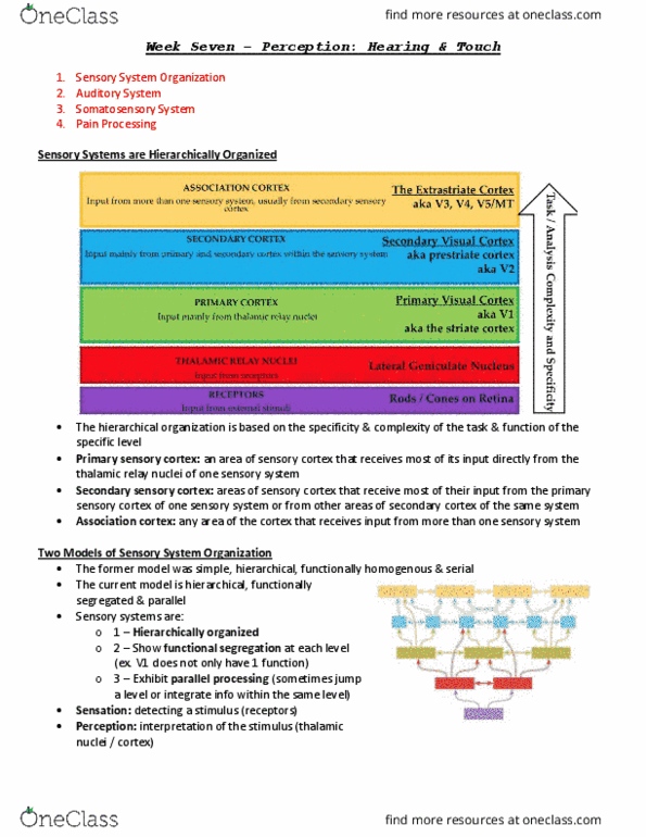
a. IN a sensitive body part like your lips, there are more number of
neurons in the somatosensory cortex to analyze the info than other
insensitive body parts.
b. Homonculus shows where in your brain that has more neurons to
dedicate to your body parts. (lots of lips, feet, hands).
3. Quick and slow adaption sensory receptors.
4. how sensory systems code the intensity and duration of a stimulus.
a. Higher frequency of action potentials = higher intensity. Can’t change
its amplitude.
b. Different frequencies of vibrations stimulates different areas of the
cochlea which goes to hair cells.
5. Rods and cones: functions and distributions on the retina
6. Visual signal transduction. Dark current in the photoreceptors, center
surround receptive fields of the ganglion cells, and signal transduction from
retina to the primary visual cortex.
a. Ganglion cells send neurotransmitters to turn off the next door
neighbor neurons (lateral inhibitions) in respect to the one that’s
turned on. Increases contrast.
b. With light, less neurotransmitters being sent to bipolar cells. Without
light, more neurotransmitters being sent to bipolar cells.
c. Amacrine cells: contribute to lateral inhibition.
7. The auditory system: sound process and frequency coding
Ch11-ANS
8. Adrenal medulla. Function and neurotransmitter release.
a. During sympathetic, releases NE and E, acting at neurohormones. (If
came from sympathetic system, NE is neurotransmitter; if came from
adrenal medulla, NE is a neurohormone).
9. Receptors used by sympathetic and parasympathetic receptors
10. Functions, neurotransmitters, and receptors of the autonomic system in the
cardiovascular systems.
Ch14-Cardio
11. Action potential of the heart contractile and pacemaker cells
12. Compare the mechanisms of muscle contraction of the skeletal muscle with
the cardiac muscle.
a. Cardiac muscles can vary force of contraction by varying calcium
entering the cell (calcium induced calcium release).
b. Does not tetanize.
find more resources at oneclass.com
find more resources at oneclass.com
Document Summary
Ch10-sensory **visual system*: the large vs the small receptor fields. Can"t change: different frequencies of vibrations stimulates different areas of the its amplitude. cochlea which goes to hair cells, rods and cones: functions and distributions on the retina, visual signal transduction. Increases contrast. light, more neurotransmitters being sent to bipolar cells: amacrine cells: contribute to lateral inhibition, the auditory system: sound process and frequency coding. Sa node in rhythm, and av node in rhythm, but not in respect of each other. 2 metronomes beating at different rhythms that"s at a mess together. Within themselves = rhythmic, looking at both = messy. Dissociation b/w qrs and p waves, dissociated: know the 4 valves and 4 vessels connect with the 4 chambers of the heart, pressure and volume curve, b to e = systole. Memorize: preload = edv, volume of ventricles at the end of diastole; beginning of systole.