BIOLOGY 2B03 Chapter Notes - Chapter 4: Inner Membrane, Intermembrane Space, Axoneme

17 Jun 2018
School
Department
Course
Professor

BIOLOGY 2B03 - Module 4 Lecture II
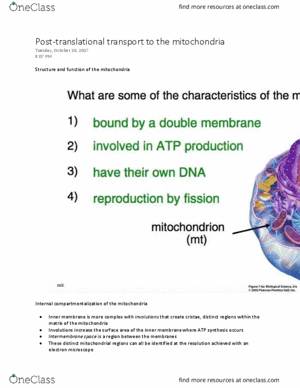
Structures and Function of the Mitochondria
● It is bound by a double membrane
○ There are interior and exterior membrane
● Mitochondria is primary site of ATP production
● The proteins of the electron transport chain found in the inner membrane
● Have their own genomes and mitochondrial genes code
● Are able to reproduce by fission
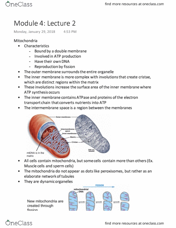
Internal compartmentalization of Mitochondria
(TEM image)
● Simple outer membrane surrounds the entire organelles
● Inner membrane is much more complex
○ Involutions that create cristae; more SA of inner membrane
■ ATP synthesis occurs here
○ Distinct regions within the matrix of the mitochondria
● Intermembrane Space: between the membranes
Sites of ATP Synthesis
● In the inner membrane: ATPase and the protein of electron transport chain convert nutrients into
cellular energy
● Elaborate structure: increase capacity of the mitochondria and create lots of ATP
More Mitochondria Provide More Energy
● Mitochondria are present in all cells but number of them vary depending on the functions of the
cells
● Cardiac Muscle: actin bundles required for continuous muscle cell contraction
○ Extensive arrays of mitochondria are providing ATP
● Sperm Cell: requires large amount of energy to maintain movement
○ The flagellar axoneme is found in the middle in cross-section, surrounded by
mitochondrias
● Neural Cell: extensive mitochondrial network
○ The nucleus, actin, and the mitochondria can be seen
● The mitochondria do not appear as punctate dots like we saw with the peroxisomes but rather as
an elaborate network of tubules
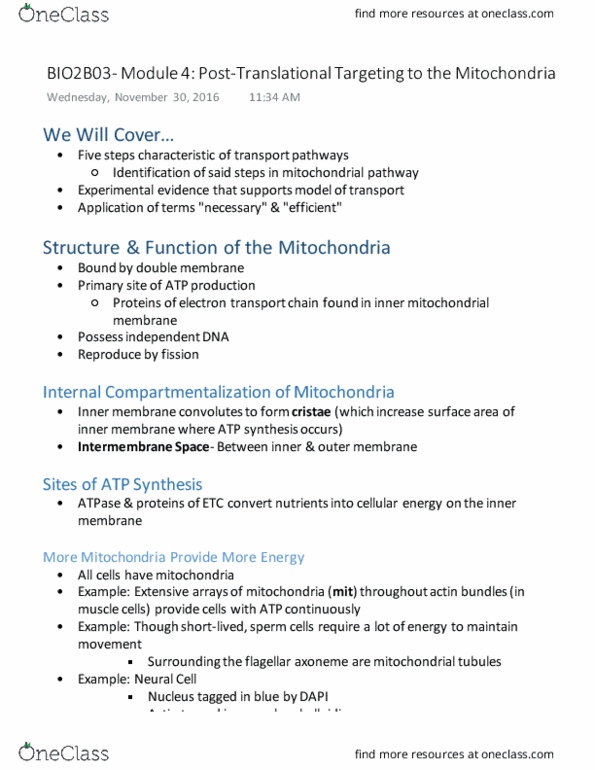
Mitochondria are dynamic and Expansive
They are very dynamic
● Able to change their shapes
● Undergo fission and fusion
● Always moving around within the cytosol
● They are moving, growing, dividing
Mitochondrial Biogenesis Requires Protein Synthesis
Mitochondria contain many mitochondrial genomes
● During fission each daughter mitochondrion usually ends up with at least one mitochondrial
genome
● No genome: will degenerate and die
● Can also grow through the import of new proteins and genesis of new membrane
find more resources at oneclass.com
find more resources at oneclass.com

Where Do Proteins Go in the Mitochondria?
Four Destination:
● The outer membrane
● The inner membrane
● The intermembrane space
● Within the matrix
Where Do Proteins Come From?
● Some proteins are coded in the mitochondrial genome and synthesized using mitochondrial
ribosomes
● Majority of proteins used in mitochondria are coded by nuclear genome
○ Theses genes are transcribed in the nucleus
○ mRNAs are translated in the cytosol by free ribosomes
○ Then proteins are transported to the mitochondria in a post-translational pathway
Is There Post-Translational Transport to Mitochondria?
Hypothesis: Fully transported proteins can be transported into the mitochondria using a post-translational
mechanism
● Two test tubes: one with energized mitochondria and protease and the other one with just the
protease
○ Protease: an enzyme that will degrade proteins in solution
● Result: the protein was protected by mitochondria from protease in the first test tube
● Why? Because the proteins have been transported into the mitochondria
● Suggestion: The fully translated protein can be transported into the mitochondria
In order to assay for protein degradation in this experiment…
● Use antibody to the mitochondrial protein
○ See whether or not that protein was localized within the mitochondria
● Alternative method: run these protein on the gel
○ If the proteins are protected, it will result in a specific band
5 Rules of Protein Transport
● There must be a signal sequence
● A receptor for that signal sequence
● A translocation channel across the membrane
● A source of energy; ATP hydrolysis in this case
● A way of targeting proteins to different locations within an organelle
RULE 1: Is There a Signal Peptide Sequence?
Matrix-targeting Motif
● Found at N-terminus
● An 18 to 50 amino acid long peptide that forms an alpha-helix
○ Specifically it is an amphipathic helix: hydrophobic on one side and hydrophilic on the
other
● Primary Structure of this Motif: Regular arrangement of hydrophobic and hydrophilic amino acid
residues
○ Residues will end up on opposite surfaces once folded into the alpha helix
Is the targeting motif “necessary” for protein transport into the mitochondria?
find more resources at oneclass.com
find more resources at oneclass.com
Document Summary
Mitochondria is primary site of atp production. The proteins of the electron transport chain found in the inner membrane. Have their own genomes and mitochondrial genes code. Simple outer membrane surrounds the entire organelles. Involutions that create cristae; more sa of inner membrane. Distinct regions within the matrix of the mitochondria. In the inner membrane: atpase and the protein of electron transport chain convert nutrients into cellular energy. Elaborate structure: increase capacity of the mitochondria and create lots of atp. Mitochondria are present in all cells but number of them vary depending on the functions of the cells. Cardiac muscle: actin bundles required for continuous muscle cell contraction. Extensive arrays of mitochondria are providing atp. Sperm cell: requires large amount of energy to maintain movement. The flagellar axoneme is found in the middle in cross-section, surrounded by mitochondrias. The nucleus, actin, and the mitochondria can be seen.