BIOLOGY 2B03 Chapter 1: BIO2B03 - Module 1 Lecture II

17 Jun 2018
School
Department
Course
Professor

BIOLOGY 2B03 - Module I Lecture II
Secondary Structures
Periodic folding of the polypeptide chain into distinct conserved,
geometric arrangements.
- Alpha-helix: spiral, rod-like structure
- Beta-sheet: planar structure, composed of alignments of two or
more beta strands
- Turns/Loops: Connectors
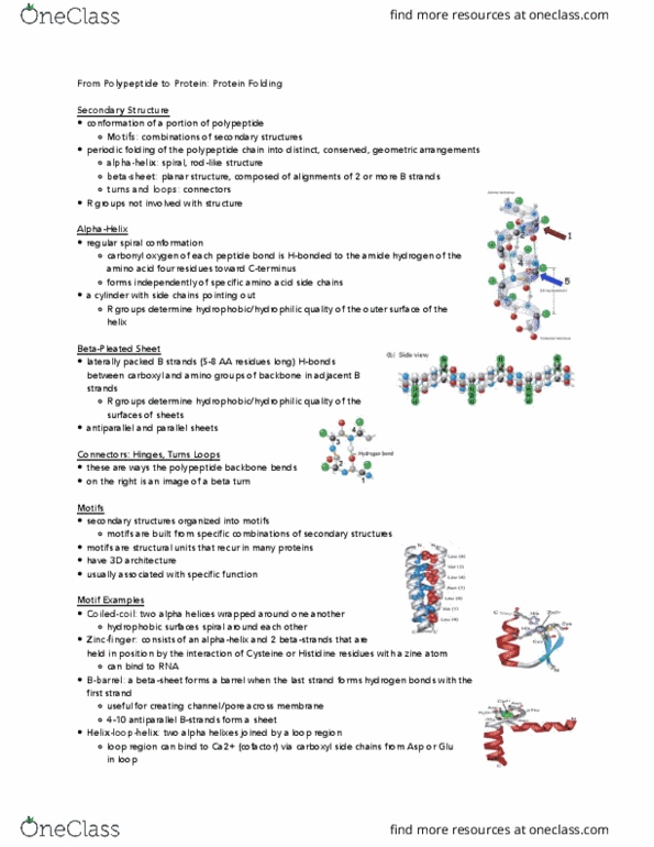
Alpha-Helix
Regular Spiral Conformation:Carbonyl oxygen of each peptide bond is
H-bonded to the amide hydrogen of the amino acid four residues toward
the C-terminus
- Forms independently of specific amino acid side chains
A Cylinder with side chains pointing out: R groups determine hydrophobic / hydrophilic quality of the outer
surface of the helix
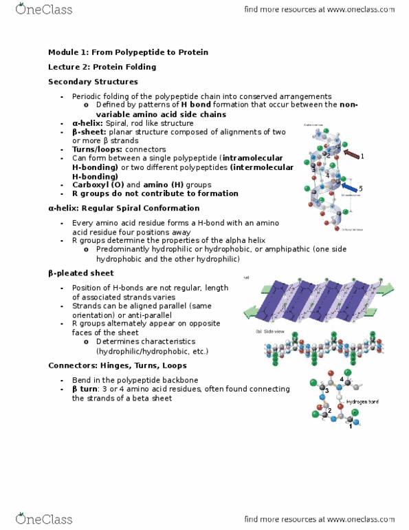
Beta-Pleated Sheet
Laterally packed beta strands (5 to 8 amino acid residues lng) H-bonds between carboxyl and amino groups of
backbone in adjacent strands
Side chains point top and bottom: R groups determine hydrophobic / hydrophilic quiality of the surfaces of the
sheets.
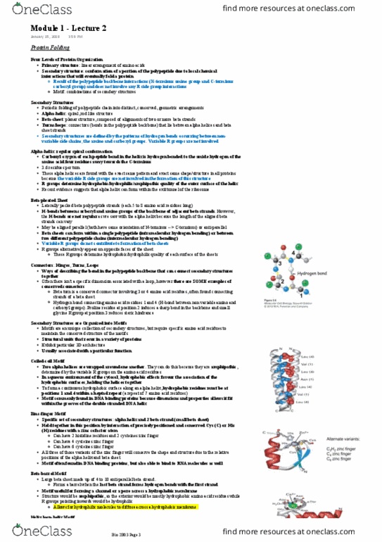
Connectors
Secondary Structures are Organized into Motifs
- Motifs are built from particular combinations of secondary structures
- Motifs are structural units that recur in a variety of proteins
- Exhibit particular 3-dimensional architecture
- Usually associated with a particular function
Coiled-Coil Motif: two alpha helices are wrapped around one another due to its amphipathic characteristic.
- Hydrophobic effects favour the association of the hydrophobic surface holding helicase together.
- Amino acid residues must be at position 1 and 4 within a heptad repeat.
- Hydrophobic surface spirals around each helix:Leucine Zipper; shows the patter of Leu-X6-Leu-X6-
Leu-X6 (X6=ny 6 amino acid)
- Commonly found in DNA binding proteins; allows it to fit within the frroces of the double stranded
DNA helix
find more resources at oneclass.com
find more resources at oneclass.com

Zinc-finger Motif: consists of an alpha-helix and two beta-strands, held in
position by the interaction of precisely positions Cys (C) or HIs (H) residues
with a zinc atom.
- Alternative variants: C2H2 Zinc finger, C4 zinc finger, C6 zinc finger
- Commonly found within the DNA binding proteins to bind to RNA
molecules as well
- Have a conserved shape and position for the alpha helix and the beta
sheet.
B-Barrel Motif: A beta-sheet forms a barrel when the last beta strand forms
hydrogen bonds with the first strand
- Loops back upon itself
- A collection of 4 and 10 anti parallel beta strands form a sheet
- Useful forming a channel or a pore across a hydrophobic membrane;
in this case the structure would be amphipathic
- Exterior: hydrophobic, interior: hydrophilic
- More hydrophilic molecules to diffuse across a hydrophobic
membrane.
Helix-Loop-Helix Motif: Two small alpha-helices joined by a loop region. Loop
region can bind Ca2+(cofactor) via carboxyl side chains from Asp or Glu in the
loop
- Can be established only once polypeptide is interacting with the calcium
- Its function depends on the cofactor.
Tertiary Structure
three dimensional arrangement of all amino acid residues of a single polypeptide
- The overall conformation of a single polypeptide
- Fundamental unit of the tertiary structure of a protein is the domain
A domain is a substructure produced by any part of a polypeptide chain that can fold independently
into a compact, single structure
Protein Domains
Functional Domains: regions of a protein that perform a certain activity
- E.g. DNA binding, enzymatic, protein-protein interaction
Structural Domains: regions of protein that forms compact, largely independent globular domains
- Proline-rich, acidic domain
find more resources at oneclass.com
find more resources at oneclass.com
Document Summary
Periodic folding of the polypeptide chain into distinct conserved, geometric arrangements. Beta-sheet: planar structure, composed of alignments of two or more beta strands. Regular spiral conformation:carbonyl oxygen of each peptide bond is. H-bonded to the amide hydrogen of the amino acid four residues toward the c-terminus. Forms independently of specific amino acid side chains. A cylinder with side chains pointing out: r groups determine hydrophobic / hydrophilic quality of the outer surface of the helix. Laterally packed beta strands (5 to 8 amino acid residues lng) h-bonds between carboxyl and amino groups of backbone in adjacent strands. Side chains point top and bottom: r groups determine hydrophobic / hydrophilic quiality of the surfaces of the sheets. Motifs are built from particular combinations of secondary structures. Motifs are structural units that recur in a variety of proteins. Coiled-coil motif: two alpha helices are wrapped around one another due to its amphipathic characteristic.