PSYCH 2E03 Chapter Notes - Chapter 4: Parahippocampal Gyrus, Extrastriate Body Area, Ocular Dominance

12 Jun 2013
School
Department
Course
Professor
Document Summary
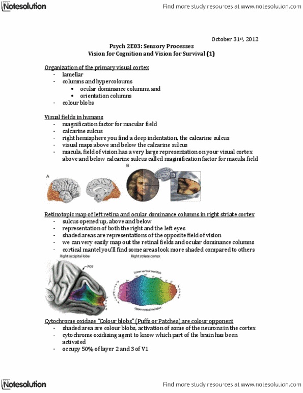
Retinotopic map: map in which each point on the lgn corresponds to a point on the retina. Receptive fields of neurons close to each other along the electrode track had receptive fields that were close to each other or overlapping on the retina. Cortical magnification factor: apportioning the small fovea with a large area on the cortex. Extra cortical space allotted to the foveal cones is available for the extra neural processing needed to accomplish high-acuity tasks. Columns: organizing for location, orientation, and ocular dominance. Ipisilateral eye: eye on the same side of the body as the lgn sends to layers 2, 3, and 5. Contralateral eye: eye on the opposite side of the lgn sends to layers. Each eye sends half of its neurons to the lgn located in the left hemisphere and half to the right. Because electrodes encounter neurons with overlapping receptive fields, neurons all receive information from the same place on retina.