HSM 330 Chapter Notes - Chapter 19: Reticular Formation, Rapid Eye Movement Sleep, Sleep Deprivation

2 Apr 2013
School
Department
Course
Professor
Document Summary
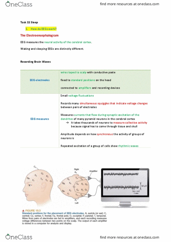
The electroencephalogram (eeg) is a measurement that enables us to glimpse the generalized activity of the cerebral cortex. The electrical contribution of any single cortical neuron is exceeding small, however, and must penetrate several layers of non-neural tissue to reach the electrode. So it takes many thousands of underlying neurons, activated together, to generate an. However, when each cell receives the same amount of excitation, but spread out in time, the summed signals are meagre and irregular. The total amount of excitation may not have changes, only the timing of the activity: an alternative way to record the rhythms is with magnetoencephalography (meg). The capabilities of meg complement those of other methods that measure brain function. It is better than eeg at localizing the sources of neural activity in the brain. Meg can record rapid fluctuations of neural activity but cannot provide the spatially detailed images of fmri.