NURS 2003H Chapter Notes - Chapter 14: Leukocyte Extravasation, Cardiac Muscle, Pus

26 Jun 2018
School
Department
Course
Professor

1
Chapter 14 –Inflammation & Wound Healing (p.248-260)
Cell Injury
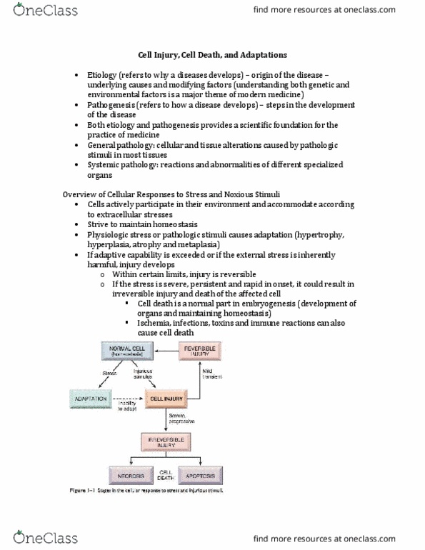
-Sublethal injury: alters function without causing cell death, potentially reversible.
-Lethal injury: irreversible process that causes cell death.
Cell Adaptation to Sublethal Injury
Hypertrophy: an increase in the size of cells, which results in increased tissue mass without cell division.
-Caused by the response of an organ or a select area of tissue to an increased demand for work.
-2 types of hypertrophy: physiological and pathological.
oPhysiological: Muscle hypertrophy results from an increase in the size of muscle fibers in
response to an increase in cellular protein, which occurs during weight training.
Other examples are uterus enlargement during pregnancy from hormonal stimulation &
enlargement of the sex organs during puberty.
oPathological: Enlargement of the heart in a person with severe hypertension to compensate for the
increased resistance to its pumping action.
Removal of one kidney results in an increase in the size of the remaining kidney because
of the increased work demand.
Hyperplasia: an increase in the # of cells because of increased cellular division.
-Reversible when the stimulus for cellular division is removed.
-Compensatory hyperplasia adaptive process whereby cells of certain organs regenerate (i.e. the liver).
-Hormonal hyperplasia occurs primarily in structures responsive to estrogen, such as breasts and the
uterus (i.e. lactation and pregnancy).
-Pathological hyperplasia an example is endometrial hyperplasia which is caused by excessive hormone
stimulation (high amounts of estrogen or an imbalance between estrogen and progesterone leads to
excessive growth of the endometrium).
Atrophy: a decrease in the size of a tissue or organ caused by a reduction in the # or the size of the individual cells.
-Frequently occurs as the result of…
oDisease (i.e. musculoskeletal disease)
oLack of blood supply (i.e. thrombus formation)
oNatural aging process (i.e. atrophy of ovaries after menopause)
oInactivity (i.e. decreased muscle size)
oNutritional deficiency
Metaplasia: the reversible transformation of one cell type into another.
-Physiological metaplasia the change of circulating monocytes to macrophages as they migrate into
inflamed tissues.
-Pathophysiological metaplasia the change of normal pseudostratified columnar epithelium of the bronchi
to squamous epithelium in response to chronic cigarette smoking.
oIf the irritating stimulus (the smoke) is removed, the bronchial metaplasia may be reversible.
Dysplasia: an abnormal differentiation of dividing cells that results in changes in the size, shape, and appearance of
these cells.
-Minor dysplasia is found in some areas of minor inflammation.
-Potentially reversible if the stimulus for the change is removed.
-Frequently a precursor of malignancy, as in cervical dysplasia.
Anaplasia: cell differentiation to a more immature or embryonic form.
find more resources at oneclass.com
find more resources at oneclass.com

2
-Malignant tumors are often characterized by anaplastic growth.
Causes of Lethal Cell Injury
-Deterioration of the nucleus…
oSuch as pyknosis –nuclear condensation and shrinkage
oKaryolysis –dissolution of nucleus and contents
oDisruption of cell metabolism
oRupture of the cell membrane
-Microbial invasion often results in cell injury and death.
-Infection occurs when pathogens (microorganisms capable of producing disease) invade and multiply in
body tissue.
TABLE 14-1 p.250
Cause Effect on Cell
Hypoxia or ischemic injury Compromised cell metabolism, acute or gradual cell death
Physical agents
-Heat
-Cold
-Radiation
-Electro thermal injury
-Mechanical trauma
Denaturation of protein, acceleration of metabolic reactions.
Decreased blood flow from vasoconstriction, slowed metabolic reactions,
thrombosis of blood vessel, freezing of cell contents that forms crystals and
can cause cell to burst.
Alteration of cell structure or enzyme systems, mutations
Interruption of neural conduction, fibrillation of cardiac muscle, coagulative
necrosis of skin and skeletal muscle.
Transfer of excess kinetic energy to cells, causing rupture of cells, blood
vessels, tissue; examples include the following…
-Abrasion: scrape of skin or mucous membranes
-Laceration: severing of vessels and tissue
-Contusion: bruise, crushing of tissue cells causing hemorrhage into
skin.
-Puncture: piercing of body
-Incision: surgical cutting.
Chemical Injury Alteration of cell metabolism, interfere with normal enzymatic action within
cells.
Microbial injury
-Viruses
-Bacteria
Taking over the cell metabolism synthesis of new particles that may cause cell
rupture; cumulative effect may produce clinical disease
Destruction of cell membrane for nucleus, production of lethal toxins
Immunological
-Antigen-antibody
response
-Autoimmune
Release of substances (histamine, complement) that can injure and damage
cells.
Activation of complement, which destroys normal cells and produces
inflammation.
Neoplastic growth Cell destruction from abnormal and uncontrolled cell growth.
Normal substances (i.e.
digestive enzymes, uric acid)
Release into abdomen, causing peritonitis and crystallization of excess
accumulation in joints and renal tissue.
Cell Apoptosis and Necrosis
-The two fundamental types of cell death
-Apoptosis: programmed death (normal event in the GI system and skin)
-Necrosis: death of a tissue (cell death on a large scale) or part of the tissue with a cellular reaction to the
dead cells.
o Dry gangrene refers to the dry shriveled and darkened area
find more resources at oneclass.com
find more resources at oneclass.com

3
o wet gangrene refers to liquefied necrotic tissue
TABLE 14-2 p.251
Defence Against Injury
-the body has various defence mechanisms to protect against injury and infection.
Mononuclear Phagocyte System
-Consists of monocytes and macrophages and their precursor cells
-Phagocytic cells are either fixed or free (mobile).
oThe macrophages of the liver, spleen, bone marrow, lunges, lymph nodes, and NS (microglial
cells) are fixed phagocytes.
oThe monocytes in the blood and the macrophages found in the connective tissue (histiocytes) are
mobile or wandering phagocytes.
-Monocytes and macrophages originate in the bone marrow.
-Monocytes spend a few days in the blood and then enter tissues and change into macrophages.
oTissue macrophages are larger and more phagocytic than monocytes.
-Functions of this system include…
oRecognition and phagocytosis (aka ingestion) of foreign material such as microorganisms
oRemoval of old damaged cells from circulation
oParticipation in the immune response
Inflammatory Response
-Complex, nonlinear process and is a key aspect of many disease processes
- It is a biological response to cell injury by pathogens, irritants, or chronic health conditions.
- It neutralizes and dilutes the inflammatory agent, removes necrotic materials, and establishes an
environment suitable for healing and repair.
- Infection almost always cause inflammation but not all inflammations are caused by infection.
-The inflammatory response can be divided into a vascular response, a cellular response, formation of
exudate, and healing.
Vascular response
oArterioles in the area briefly undergo transient vasoconstriction, which is stimulated by the SNS.
oPlatelets adhere to vessels and aggregate to seal the injured area, forming a fibrin-platelet clot, and
release proinflammatory mediators such as histamine which cause vasodilation.
oThis results in hyperemia (increased blood flow) in which filtration pressure increases, causing
endothelial cell retraction and increase in capillary permeability.
oMovement of fluid from the capillaries into tissue spaces is thus facilitated.
Initially composed of serous fluid, this inflammatory exudate later contains plasma
proteins, primarily albumin, which exerts oncotic pressure that further draws fluid from
blood vessels, and the tissue becomes edematous.
oPlasma protein fibrinogen leaves the blood and is activated by the products of the injured cells to
become fibrin.
Fibrin strengthens the blood clot formed by platelets.
Clot traps bacteria, prevents their spread, and serves as a framework for the healing
process.
Cellular response
oPhagocytes produce nitric oxide
find more resources at oneclass.com
find more resources at oneclass.com
Document Summary
Sublethal injury: alters function without causing cell death, potentially reversible. Lethal injury: irreversible process that causes cell death. Hypertrophy: an increase in the size of cells, which results in increased tissue mass without cell division. Caused by the response of an organ or a select area of tissue to an increased demand for work. 2 types of hypertrophy: physiological and pathological: physiological: muscle hypertrophy results from an increase in the size of muscle fibers in response to an increase in cellular protein, which occurs during weight training. Removal of one kidney results in an increase in the size of the remaining kidney because of the increased work demand. Hyperplasia: an increase in the # of cells because of increased cellular division. Reversible when the stimulus for cellular division is removed. Compensatory hyperplasia adaptive process whereby cells of certain organs regenerate (i. e. the liver).