PSYCH312 Chapter Notes - Chapter 10: Functional Magnetic Resonance Imaging, Occipital Lobe, Temporal Lobe

19 Nov 2015
School
Department
Course
Professor
Document Summary
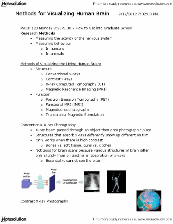
Medical aspects of learning disabilities and related mild disabilities (ch. 10): Recent brain research: dyslexia (interferes with learning to read) Related to brain structure + function (not a matter of intelligence) Postmortem anatomical studies: analyzed brain tissue of deceased individuals who had dyslexia, abnormality in planum temporale. Superior surface of temporal lobe (center of language control) Left hemisphere (had fewer brain cells than that of nondyslexic individuals) Right hemisphere (same area was larger + contained more cells than found in nondyslexic individuals) Computed tomography: computerizd series of x-rays that build a 3d image of the brain. Positron-emission tomography (pet): measures blood flow to the brain regions through use of radioactive compound that is injected into the bloodstream. Rely heavily on the front of the brain (phoneme producer region) when they try to say, the sounds of phonemes. Word analyzer region (as they try to decode words)