ANP 1106 Chapter Notes -Cerebral Cortex, Spiral Ganglion, Thalamus

21 Jan 2013
School
Department
Course
Professor
Document Summary
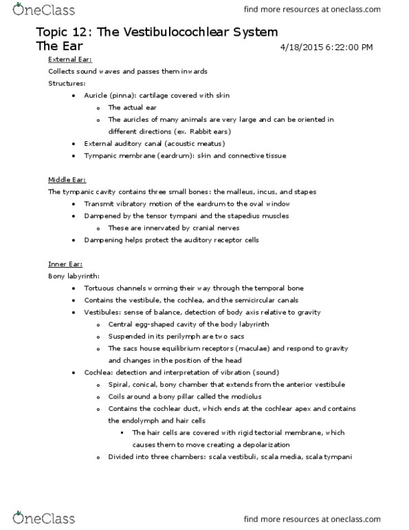
Divided into external ear, middle ear, and internal ear. The external ear and middle ear structures are involved with hearing only and are simple. The internal ear functions in both equilibrium and hearing and is complex. Consists of the auricle and the external acoustic meatus. The auricle (pinna) is the shell-shaped projection surrounding the opening of the external acoustic meatus. Its outer rim is known as the helix. The lobule (earlobe) is dangling and lacks supporting cartilage. Functions to direct sound waves into the external acoustic meatus. The external acoustic meatus (auditory canal) is a short, curved tube that extends from the auricle to the eardrum. The canal is lined with skin bearing hairs, sebaceous glands, and ceruminous glands that secrete cerumen which traps foreign particles and repels insects. Sound waves entering the external acoustic meatus eventually hits the tympanic membrane (eardrum) which acts as the boundary between the outer and middle ears.