PSY280H5 Chapter Notes - Chapter 5: Ocular Dominance Column, Optic Chiasm, Magnocellular Cell

Document Summary
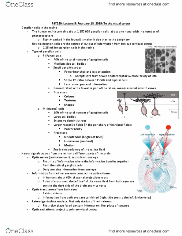
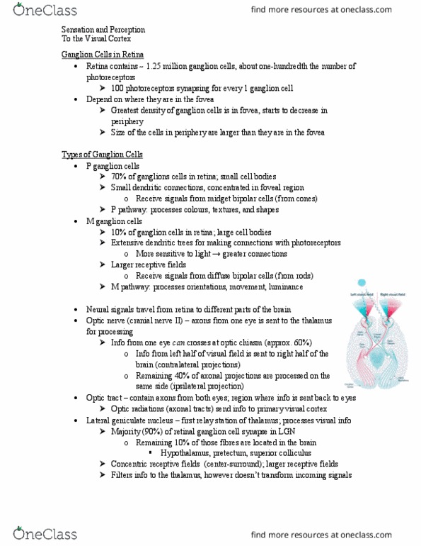
Describe how retinal ganglion cells cross over in the optic chiasm. Where the optic nerve from each eye meet together behind the eyes. Axons from the ganglion cells from the right half and left retina and right half of right retina combine forming the right optic tract, proceeds to the right hemisphere of the brain. Left lgn receives input from right visual world. Right lgn receives input from the left visual world. Magnocellular: sensitive to motion, light detection, sudden changes in visual image. ----- > parasol retinal ganglion cells project to magnocellular layer of lgn. ------ > midget ganglion cells project to parvocellular layer of lgn. ------ > bistratified retinal ganglion cells project to koniocellular layer of lgn. Allocation of more space in the cortex to some sensory receptors. Fovea is less than 1% of retina but takes up 50% of v1, fovea specializes in color and visual acuity, most important in determining what an object is.