BIOB10H3 Chapter Notes - Chapter 18 (696-700, 711-713, 715-716, 737-740): Green Fluorescent Protein, Fluorophore, Differential Centrifugation

Document Summary
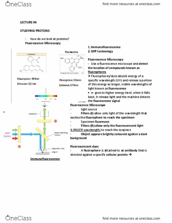
The fluorescence microscopy allows for observation of certain molecules and their location called fluorophore. Fluorescence is called when the fluorophore absorbs photon of a specific wavelength and release energy in longer wavelength. Light is beamed into the fluorescence microscopy, and this light travels through a filter that blocks all wavelength from travelling except those that excite the fluorophore. The light that is focused on the fluorophore is excited and it emits visible wavelength that can be seen through the objective lens. Immunofluorescence is a technique used when covalently linking organic fluorophore to an antibody to determine the location of a specific protein. Fluorescence can be used to determine the location of dna and rna and their specific nucleotide bases sequences, they can be used to study the transmembrane protein, and protein in dynamic motion. In most cases, fluorescence has been made by conjugating chemically with fluorophore; in some other cases, like jelly fish, there are green fluorescent protein (gfp).