PSYB64H3 Chapter Notes - Chapter 2: Motor System, Diencephalon, Motion Sickness

Document Summary
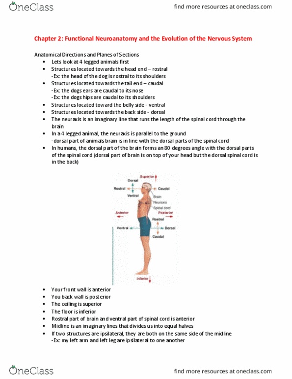
The anatomy and evolution of the nervous system. The following are for 4 legged animals: the neuraxis forms a straight line running parallel to the ground. Rostral or anterior = structures that are located toward the head end of the animal: ex. Head of the dog is rostral to its shoulders. Caudal or posterior = structures located toward the tail end of the animal: ex. The dog"s ears are caudal to its nose and its hips are caudal to its shoulders. Inferior or ventral = structures located toward the belly side. Superior of dorsal = structures towards the back. The dorsal parts of the animal"s brain are in line with the dorsal parts of the spinal cord. For people, the directions are different because our 2 legged stance puts a 90 degree bend in the neuraxis (an imaginary line that runs the length of the spinal cord through the brain)