MCD BIO 138 Chapter Notes - Chapter lec 16: Trachea, Gastrointestinal Tract, Colorectal Cancer

13 Oct 2020
School
Course
Professor
Lecture 16. DEVELOPMENT OF THE ENDODERMAL DERIVATIVES IN VERTEBRATES
1. Left-right asymmetry
Vertebrates are bilaterally symmetric about the midline for many structures, like eyes,
nasal passages, and limbs, but most internal organs are arranged asymmetrically. For example,
the heart is on the left side, and left lung has fewer lobes than the right lung. Similarly, the
endodermal organs exhibit invariant asymmetric placement within the abdominal cavity, and this
is required in order to provide enough space for each organ. Thus, while all of the internal
organs display some asymmetry in their placement along the L/R axis, this asymmetry is easy
to see with respect to endodermally-derived organs, which we will discuss below.
L/R asymmetry is controlled early in development, and the node is a crucial signaling
center for specifying identity along the L/R axis. In birds and mammals, cilia located in the
node are required for L/R asymmetry. How do we know this? Mutations in left-right dynein (lrd)
cause randominzation of organ placement. Recall that dyneins are motor proteins. In addition to
moving cargos along microtubules, they are also required for cilia to beat. Other evidence that
cilia are involved in L/R asymmetry comes from humans with Kartagener’s syndrome. In these
individuals, organ placement becomes random; this causes problems as a consequence of
organ crowding. In these individuals, all cilia are immotile. Therefore, these individuals have
respiratory defects (cells in the respiratory tract are ciliated) and males are infertile (the sperm
flagellum can’t rotate). Mutations in lrd are found in some individuals with Kartagener’s
Syndrome. Other individuals with Kartagener’s Syndrome have mutations in different ciliary
proteins. It is not entirely clear how cilia are involved in generating asymmetry, but it is known
that the cilia in the node all rotate in the same direction, and this causes a directional fluid flow
across the node. Thus, this directional flow could lead to localization of a determinant or
activation of a signaling pathway on one side of the node compared to the other. This differential
activity subsequently leads to differential activation of gene expression in the mesoderm on the
right and left sides of the embryo.
2. Embryonic morphogenesis of the endoderm and its derivatives
2.1. Specification and regionalization of endoderm
After gastrulation, the endoderm represents the innermost germ layer. In lower
vertebrates, it is a hollow tube composed of a single layer of epithelial cells that will give rise to
the future gut, lungs, liver, pancreas, and other structures. This epithelial tube is surrounded by
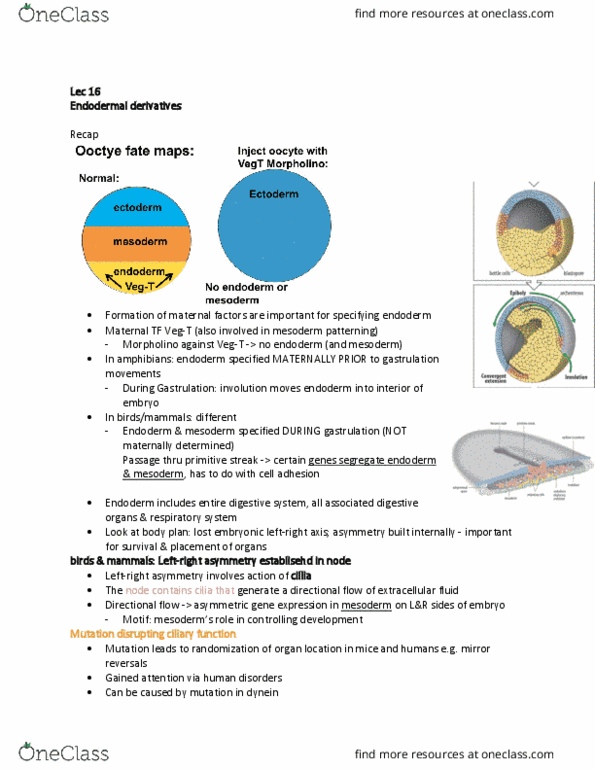
mesenchyme of mesodermal origin. As discussed previously, in amphibians, the vegetal cells
are specified as endoderm due to the presence of maternal factors such as VegT.
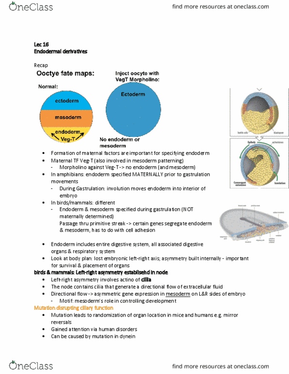
In birds and mammals, the early embryo is flat. The endodermal and mesodermal germ
layers in birds and mammals are specified as cells ingress through the primitive streak. The
endodermal cells form a layer underneath the mesoderm. This flat embryo transforms into a
closed cylinder. To achieve this, the newly formed endoderm has to fold. The folding occurs as
active invagination processes initiated at the anterior and posterior ends of the endoderm.
Following closure, the endoderm forms an epithelial cylinder surrounded by a thin layer of
mesenchymal mesodermally-derived cells that differentiate into smooth muscle. This
invagination of the endoderm and associated mesenchyme transforms the initially flat bird and
mammalian embryo into a tubular structure.
Regional differences in epithelial cell shapes and cell proliferation result in the
demarcation of three endodermal segments, the foregut, midgut, and hindgut. in mammals, the
foregut gives rise to the pharynx, esophagus and stomach. The lung grows out from the ventral
side of the esophagus. The midgut develops into small intestine; liver and pancreas originate as
outgrowths from the midgut. The hindgut forms the large intestine and colon.

2.2. Development of endodermal derivatives
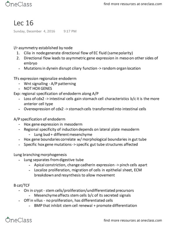
Many endodermal organs develop as invaginations from the gut tube. For example, the
lung bud invaginates from the foregut. The lung bud then bifurcates to form two buds, which will
become the right and left lungs. The bronchial buds elongate and bifurcate (branch) further,
ultimately creating a large surface area (which equals a large football field in size). This process
is known as branching morphogenesis. Each branch ends in a small sac-like structure called
an alveolus. Capillaries form a network around each alveolus, which thereby become the
structures responsible for gas exchange.
Similar to the lung bud and its branches, the hepatic (liver) and pancreatic buds are
invaginations of the endoderm, which, after profuse branching, give rise to the liver and
pancreas. Depending on the endodermal organ, the cells in the branched epithelium will
differentiate along specialized pathways. For example, in the pancreas, the branched
endodermal epithelium differentiates into exocrine glands that produce enzymes that are
excreted into the small intestine where they are essential for digestion. Interspersed within the
exocrine pancreatic glands are endocrine glands, called islet of Langerhans (or islets, for short).
Islet cells produce the hormones insulin and glucagon.
The stomach, intestines (large and small) and rectum develop from the gut tube. Under
the influence of molecules that provide A/P identity, the cells in the appropriate regions of the
gut tube differentiate into cell types that carry out the distinct functions of these organs.
3. Molecular mechanism controlling regionalization of the endoderm
3.1 Specification of the endoderm
As discussed above, in amphibians, maternal factors such as VegT specify the
endoderm. In mammals, the endoderm is specified as cells move through the primitive streak.
In Xenopus, the endoderm and mesoderm involute through the dorsal blastopore lip together,
and are patterned along the dorsal/ventral axis by the same signaling molecules. Similarly, in
birds and mammals, the mesoderm and endoderm ingress together through the primitive streak.
3.2. Reciprocal interactions between endoderm and mesoderm
Interactions between endoderm and the surrounding mesoderm play a crucial role in the
development of endodermal organs. Classical experiments have shown that endodermal cells
isolated from the surrounding mesoderm and cultured in vitro do not differentiate. If the
mesoderm that surrounds the gut tube is co-cultured along with the endoderm, organ specific
differentiation occurs. Furthermore, the type of organ that develops depends on the mesoderm,
not the endoderm, along the antero-posterior axis. For example, if tracheal bud endoderm is co-
cultured with mesoderm from different regions, it differentiates according to the location from
where the mesoderm is derived. Mesoderm from near the liver leads to the formation of liver-
like tubules in the tracheal bud. This regional specificity of induction controlled by the
mesoderm was seen previously when we considered the development of cutaneous structures.
Mesoderm outside the immediate layer surrounding the endoderm can also play an instructive
role in the specification of some endodermal derivatives. For example, signals from the
notochord (which lies dorsal to the endodermal gut tube) and the heart primordium (ventral to
the endodermal gut tube) play an essential role in the specification of the pancreas and liver on
the dorsal and ventral surfaces of the gut tube, respectively. Inductive signals also pass from
the endoderm to the mesoderm. In other words, signaling between the mesoderm and
endoderm is reciprocal.
3.3. Role of Hox genes and other organ-specific regulatory genes
The ParaHox complex consists of three homeodomain-containing genes, and
these play a key role in specifying A/P identity along the gut tube. Like the genes of the Hox
complex, the three homeobox genes of the ParaHox complex (Cdx, Pdx, and Nkx) are
Document Summary
L/r asymmetry is controlled early in development, and the node is a crucial signaling. Vertebrates are bilaterally symmetric about the midline for many structures, like eyes, Development of the endodermal derivatives in vertebrates: left-right asymmetry nasal passages, and limbs, but most internal organs are arranged asymmetrically. For example, the heart is on the left side, and left lung has fewer lobes than the right lung. Similarly, the endodermal organs exhibit invariant asymmetric placement within the abdominal cavity, and this is required in order to provide enough space for each organ. In birds and mammals, cilia located in the node are required for l/r asymmetry. Mutations in left-right dynein (lrd) cause randominzation of organ placement. In addition to moving cargos along microtubules, they are also required for cilia to beat. Other evidence that cilia are involved in l/r asymmetry comes from humans with kartagener"s syndrome. In these individuals, organ placement becomes random; this causes problems as a consequence of organ crowding.