PHYSIOL 335 Chapter 9, 12: Physiology 335 Lab 6 Reading notes

28 Oct 2016
School
Department
Course
Professor
Document Summary
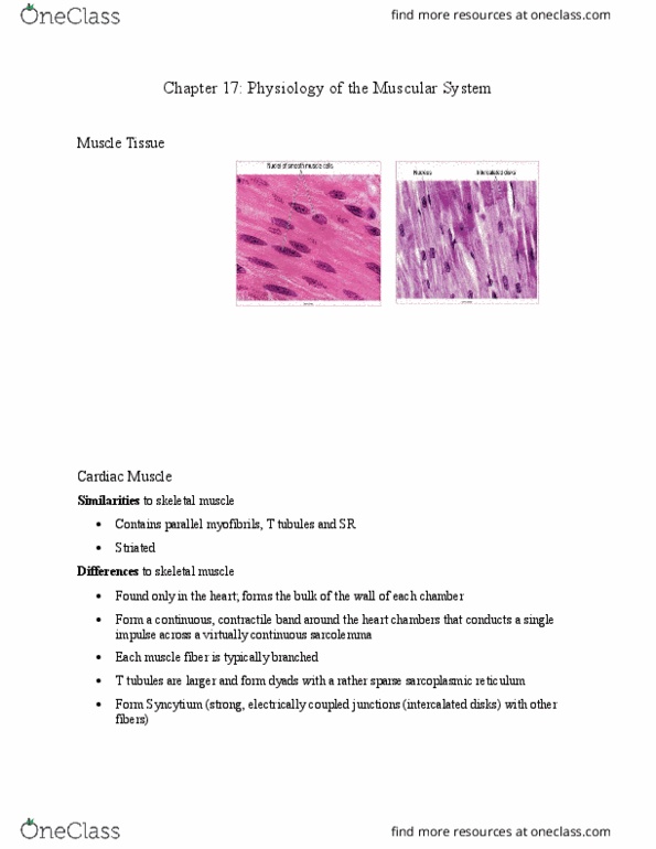
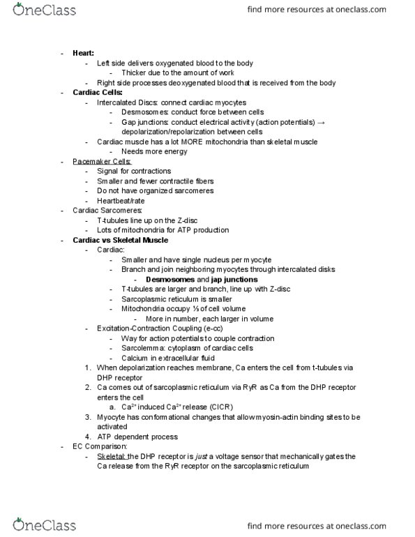
Lab 6: cardiovascular physiology (ecg) and syphgmomamometry reading. Cellular structure of cardiac muscle: striated due to repeating sarcomeres (sarcomeres made of repeating thin and thick filaments, troponin and tropomyosin present in the thin filament. Functions the same as in skeletal muscle: contains t tubules and a ca2+ loaded sarcoplasmic reticulum. Mechanism by which these membranes interact to release ca2+ is different than in skeletal muscle: single nucleus, intercalated disks join adjacent cells together. Gap junctions also found here help the heart to work as a unit. Ec coupling in cardiac muscle: depolarization during cardiac muscle cell aps is in part due to an influx of ca2+ through specialized voltage-gated channels called l-type ca2+ channels (l = long lasting current) L type channels are a type of dhp receptor: steps. Membrane is depolarized by na+ entry as an action potential begins.