PSYC10003 Lecture Notes - Lecture 3: Myasthenia Gravis, Dendritic Spine, Autoimmune Disease

13 Jun 2018
School
Department
Course
Professor

6th March ‘18
MBB1 Week 2; Lecture 3 notes
Structure and function of the human nervous system – Part 1
-Myasthenia Gravis: disorder of synaptic transmission (autoimmune disorder like MS)
-extreme fatiguability
-fluctuating muscle weakness
(proximal > distal)
-problems chewing (dysphagia) &
talking (dystharthia)
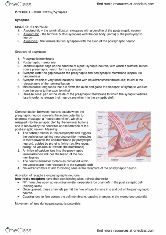
-synapse: means of communication
between neurons
-presynaptic membrane
-postsynaptic membrane
-dendritic spine
-synaptic cleft
-synaptic vesicles
-microtubules
-release zone
-release of neurotransmitter
-vesicles contain neurotransmitter (NT) molecules
-AP in pre-synaptic cell triggers vesicles to move towards cell membrane
-vesicles guided toward membrane by proteins
-guiding proteins - act like ropes - to help pull vesicle & presynaptic membrane tgt
-influx of calcium ions into presynaptic terminal button = fusion of two membranes
-neurotransmitter molecules - released into synaptic cleft
-activation of receptors on post synaptic membrane
-ionotropic receptors = own building sites
-when neurotransmitter molecule attaches to binding site = ion channel opens (like key in lock)
-ions flow thru cell membrane = change in membrane AP
-chemical signal back into electrical signal
-movement of ions
-post synaptic membrane:
find more resources at oneclass.com
find more resources at oneclass.com
Document Summary
Structure and function of the human nervous system part 1. Myasthenia gravis: disorder of synaptic transmission (autoimmune disorder like ms) Ap in pre-synaptic cell triggers vesicles to move towards cell membrane. Guiding proteins - act like ropes - to help pull vesicle & presynaptic membrane tgt. Influx of calcium ions into presynaptic terminal button = fusion of two membranes. Neurotransmitter molecules - released into synaptic cleft. Activation of receptors on post synaptic membrane. When neurotransmitter molecule attaches to binding site = ion channel opens (like key in lock) Ions flow thru cell membrane = change in membrane ap. Excitatory postsynaptic potentials depolarise postsynaptic cell membrane. Increases likelihood that ap will be triggered in postsynaptic neuron. Combined effect of epsp = determines whether this neuron has axon potential. Post synaptic membrane potential before neurotransmitter release - -70 mv (resting level) Post synaptic membrane potential after neurotransmitter release = depolarisation. Inhibitory postsynaptic potentials hyper-polarise postsynaptic cell membrane.