NURS 301 Lecture Notes - Lecture 7: Inferior Mesenteric Artery, Superior Mesenteric Artery, Lesser Omentum


Digestive System
Functions and Processes
Taking in fuel, making energy
ATP
•
-
Making building block
-
Types of digestion
Mechanical
Chewing
•
-
Chemical
Using enzymes
•
-
Digestive system functions
Ingestion: Intake of food
o
Digestion: mechanical and chemical breakdown
o
Absorption: Uptake of nutrients
o
Compaction
Absorption of water
§
Consolidation of indigestible residue
§
o
Defecation: Elimination of feces
o
General anatomy
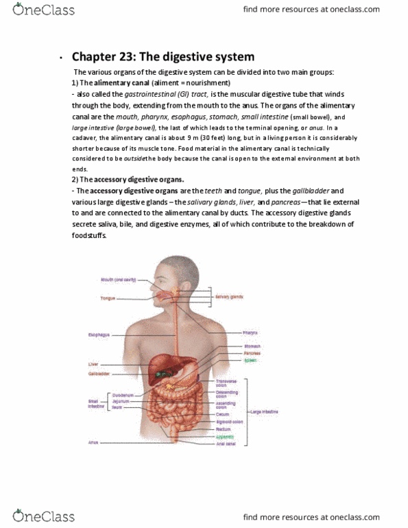
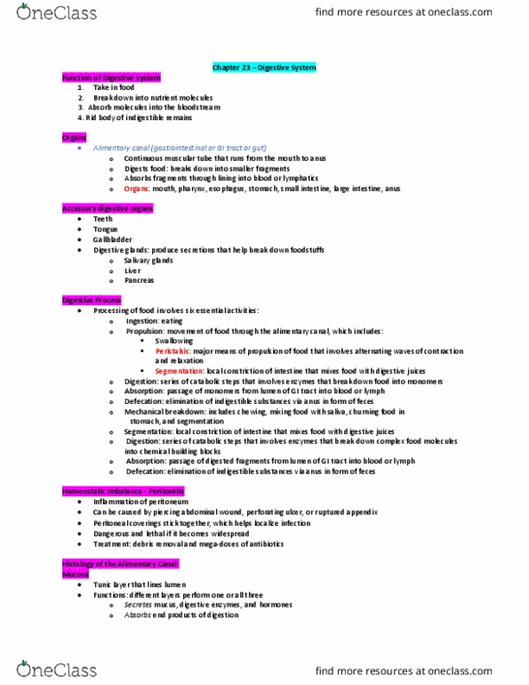
Alimentary canal
Oral cavity, anus
§
o
Gastrointestinal tract
Stomach, intestine
§
o
Accessory glands and structure
o
Alimentary Canal Wall has 4 layers
Variation occurs along the length of the GI tract; from the esophagus to
the anal canal
Mucosa
§
Submucosa
§
Muscularis
§
Serosa: outer surface
Serous membrane
§
§
-
Enteric nervous system
Nervous network to esophagus, stomach, intestines
•
Submucosal plexus
Controls gland secretions
○
•
Myenteric plexus
Found in muscularis layer
○
Peristalsis
○
•
Extensive from Autonomic NS
Sympathetic
○
Parasympathetic
Controls more of digestive tract
□
○
•
-
Blood Supply
Foregut
Esophageal arteries
•
Celiac trunk
•
-
Midgut
Superior mesenteric artery
•
-
Hindgut
Inferior mesenteric artery
•
-
Peritoneum and Mesenteries
Peritoneum is the serous membrane lining the abdominopelvic cavity
and covering the surfaces of the organs within
Visceral peritoneum = serosa
Outer membrane, covers organs
○
Lining of cavity
○
•
Parietal peritoneum = lines the cavity and is continuous with the
visceral peritoneum
•
Peritoneal cavity = space between containing serous fluid
•
Peritonitis
•
-
Mesentaries = connective tissue sheets holding abdominal viscera in
place
Double layers of serous membranes
•
Help to attach organs to the back wall
•
Function to provide blood vessels, lymphatics and nerves
•
Hold organs in place
•
Store fat, lymph nodes
Mesocolon = binds large intestine to the posterior wall
○
Lesser omentum suspends stomach and small intestine
(duodenum portion)
○
•
-
Lesser omentum = extends from lesser curvature of stomach to liver
Connects stomach to liver
•
-
Greater omentum = hangs down like an apron from stomach's greater
curvature
Drapes over the ventral surface of the viscera
•
Loaded with fat
•
Comes from inferior margin of stomach, draps over intestines
•
Goes around intestine and wraps to the back and connects to
mesocolon
•
-
Mesocolon = messentary of the colon
-
Retroperitoneal = back of the cavity
E.g. kidneys, pancreas, parts of large intestine
Oral Cavity, Pharynx and Esophagus
Oral Cavity (Buccal Cavity)
Ingestion
•
Digestion
•
Taste
•
Voice production
•
Swallowing
•
Breathing
•
-
Food's Journey Starts in Oral Cavity
Lips (labia) and cheeks: skeletal muscle covered externally by
skin; buccinators form cheeks; orbicularis oris forms lips. Inside
lining is stratified squamous cell epithelium
•
Lips and cheeks help keep food between teeth when we chew; and
aid in speech
•
The lips are large
They extend from inferior margin of the nose to the superior
border
○
•
Easier to see blood, no keratin in lips
Tissue more transparent
○
Blood flow to there is apparent
○
•
Palate
Forms the roof of mouth; hard palate anterior, soft palate
posterior (uvula hands down)
○
Soft palate is anchored to the tongue
○
•
Tongue
Occupies the floor of the mouth and fills most of the oral
cavity
Bundles of skeletal muscle = intrinsic muscles
□
Extrinsic muscles attach it to skull and hyoid bone
□
Lingual frenulum secures it to the floor
□
Mucosa covering
□
○
Covers nasal pharynx, so you don't get food into nasal cavity
○
Papillae of Tongue
Defined
□
For friction
Allow licking
®
Round pinkish
®
Taste receptors in them
®
□
Kids have a stronger sense of flavor
□
○
•
-
The Teeth
Also referred to as dentition
-
Mastication
-
2 sets: 20 deciduous vs. 32 permanent teeth
-
Incisors - chisels
-
Canines - puncture and shred
-
Premolars and molars - crush and grind
-
Embedded in mandible or maxilla
Periodontal ligament
•
Cementum of root
•
-
Gingiva (gums) surrounds neck
-
Enamel covers crown
-
Internal structures:
Dentin
•
Pulp cavity, root canal
•
-
The Salivary Glands Locations
Extrinsic salivary glands
Parotid - near ear
•
Duct opens at 2nd upper molar
•
-
Submandibular - under jaw
Ducts open at lower central incisors
•
-
Sublingual - under tongue
Several duct openings under tongue
•
-
Intrinsic salivary glands
-
Saliva
Lubricates, digestion, cleanses, teeth, inhibits microbes, dissolves
for taste
•
Contains
Amylase and lipase
○
Mucus
○
Lysozyme
○
Immunoglobulin A
○
Electrolytes (salts)
○
•
-
The Pharynx and Esophagus
Pharynx = funnel for air and food (bolus)
Oropharynx and laryngopharynx pass food
•
Walls contains skeletal muscle
•
-
Esophagus - tube transport food to stomach
Starts behind larynx
•
Passes through esophageal hiatus of diaphragms
•
Ends a lower esophageal sphincter located at cardiac orifice to
stomach
•
-
Deglutition
Swallowing
•
Coordinated by swallowing center of medulla
•
-
Gastroesophageal Reflux and Peptic Ulcer
Heart Burn
Flush of chyme comes up to esophagus, acids
•
-
Stomach, Small Intestines, and Large Intestines
The Stomach
Reservoir for food
•
Digestion
•
Chyme
•
Rugae for expansion
•
Three layers of muscle in muscularis
•
Can hold 4 liters
•
Microscopic Anatomy
Gastric pits of mucosa with glands
Mucous cells - mucus
□
Regenerative (stem) cells - make new cells
□
Parietal cells - HCl, intrinsic factor, ghrelin
□
Chief cells - pepsinogen, gastric lipase
□
Enteroendocrine cells - hormones
□
○
•
Stomach Mucosa is protected
Mucous coat
Thick
□
Highly alkaline
□
○
Tight junctions between epithelial cells prevent seepage of
gastric juice
○
Epithelial cells are frequently replaced (they only live 3-6
days)
○
Zygomens: Enzymes that digest protein are secreted in an
inactive form (pepsinogen)
○
•
-
Small Intestines
Digestion
•
Absorption of nutrients
•
Gross Anatomy
Duodenum
First 25 cm (10in)
□
Circular folds (plicae circulares)
□
○
Jejunum
1-1.7 m
□
Mostly within umbilical region
□
Most digestion and absorption occur here
□
Prominent circular folds
□
○
Ileum
1.6-2.7 m
□
Peyer patches
□
Ileocecal junction
Ileocecal valve
®
□
○
•
Intestinal Mucosa
Projections increase surface area
○
Villi, microvilli
○
Fats too big to get into blood vessels
○
•
-
Large Intestine
Bacterial fermentation
•
Water, salts, and some vitamin absorption
•
Reservoir for and removal of waste (feces)
•
Descending and Sigmoid Colon
•
Gross Anatomy
Taeniae coli - muscle
○
Haustra - pouches
○
Cecum - blind end
Appendix - blind tube
□
○
Colon
Ascending colon
On right side of body, right colic flexure
®
□
Transverse colon
Left colic flexure
®
□
○
Rectum
Transverse rectal folds (valves) retain feces
□
○
Anal canal (3 cm long)
Internal smooth muscle and external skeletal anal
sphincters
□
○
•
Microscopic Anatomy
Epithelium is mostly simple columnar
Exception: anal canal has stratified squamous
□
Has intestinal crypts
□
Abundant lymphatic tissue
□
Mucosa specialize for fluid and electrolyte (Salts)
absorption
□
○
•
-
Week 7 - 5/7
Monday, May 7, 2018
3:35 PM

Digestive System
Functions and Processes
Taking in fuel, making energy
ATP
•
-
Making building block
-
Types of digestion
Mechanical
Chewing
•
-
Chemical
Using enzymes
•
-
Digestive system functions
Ingestion: Intake of food
o
Digestion: mechanical and chemical breakdown
o
Absorption: Uptake of nutrients
o
Compaction
Absorption of water
§
Consolidation of indigestible residue
§
o
Defecation: Elimination of feces
o
General anatomy
Alimentary canal
Oral cavity, anus
§
o
Gastrointestinal tract
Stomach, intestine
§
o
Accessory glands and structure
o
Alimentary Canal Wall has 4 layers
Variation occurs along the length of the GI tract; from the esophagus to
the anal canal
Mucosa
§
Submucosa
§
Muscularis
§
Serosa: outer surface
Serous membrane
§
§
-
Enteric nervous system
Nervous network to esophagus, stomach, intestines
•
Submucosal plexus
Controls gland secretions
○
•
Myenteric plexus
Found in muscularis layer
○
Peristalsis
○
•
Extensive from Autonomic NS
Sympathetic
○
Parasympathetic
Controls more of digestive tract
□
○
•
-
Blood Supply
Foregut
Esophageal arteries
•
Celiac trunk
•
-
Midgut
Superior mesenteric artery
•
-
Hindgut
Inferior mesenteric artery
•
-
Peritoneum and Mesenteries
Peritoneum is the serous membrane lining the abdominopelvic cavity
and covering the surfaces of the organs within
Visceral peritoneum = serosa
Outer membrane, covers organs
○
Lining of cavity
○
•
Parietal peritoneum = lines the cavity and is continuous with the
visceral peritoneum
•
Peritoneal cavity = space between containing serous fluid
•
Peritonitis
•
-
Mesentaries = connective tissue sheets holding abdominal viscera in
place
Double layers of serous membranes
•
Help to attach organs to the back wall
•
Function to provide blood vessels, lymphatics and nerves
•
Hold organs in place
•
Store fat, lymph nodes
Mesocolon = binds large intestine to the posterior wall
○
Lesser omentum suspends stomach and small intestine
(duodenum portion)
○
•
-
Lesser omentum = extends from lesser curvature of stomach to liver
Connects stomach to liver
•
-
Greater omentum = hangs down like an apron from stomach's greater
curvature
Drapes over the ventral surface of the viscera
•
Loaded with fat
•
Comes from inferior margin of stomach, draps over intestines
•
Goes around intestine and wraps to the back and connects to
mesocolon
•
-
Mesocolon = messentary of the colon
-
Retroperitoneal = back of the cavity
E.g. kidneys, pancreas, parts of large intestine
Oral Cavity, Pharynx and Esophagus
Oral Cavity (Buccal Cavity)
Ingestion
•
Digestion
•
Taste
•
Voice production
•
Swallowing
•
Breathing
•
-
Food's Journey Starts in Oral Cavity
Lips (labia) and cheeks: skeletal muscle covered externally by
skin; buccinators form cheeks; orbicularis oris forms lips. Inside
lining is stratified squamous cell epithelium
•
Lips and cheeks help keep food between teeth when we chew; and
aid in speech
•
The lips are large
They extend from inferior margin of the nose to the superior
border
○
•
Easier to see blood, no keratin in lips
Tissue more transparent
○
Blood flow to there is apparent
○
•
Palate
Forms the roof of mouth; hard palate anterior, soft palate
posterior (uvula hands down)
○
Soft palate is anchored to the tongue
○
•
Tongue
Occupies the floor of the mouth and fills most of the oral
cavity
Bundles of skeletal muscle = intrinsic muscles
□
Extrinsic muscles attach it to skull and hyoid bone
□
Lingual frenulum secures it to the floor
□
Mucosa covering
□
○
Covers nasal pharynx, so you don't get food into nasal cavity
○
Papillae of Tongue
Defined
□
For friction
Allow licking
®
Round pinkish
®
Taste receptors in them
®
□
Kids have a stronger sense of flavor
□
○
•
-
The Teeth
Also referred to as dentition
-
Mastication
-
2 sets: 20 deciduous vs. 32 permanent teeth
-
Incisors - chisels
-
Canines - puncture and shred
-
Premolars and molars - crush and grind
-
Embedded in mandible or maxilla
Periodontal ligament
•
Cementum of root
•
-
Gingiva (gums) surrounds neck
-
Enamel covers crown
-
Internal structures:
Dentin
•
Pulp cavity, root canal
•
-
The Salivary Glands Locations
Extrinsic salivary glands
Parotid - near ear
•
Duct opens at 2nd upper molar
•
-
Submandibular - under jaw
Ducts open at lower central incisors
•
-
Sublingual - under tongue
Several duct openings under tongue
•
-
Intrinsic salivary glands
-
Saliva
Lubricates, digestion, cleanses, teeth, inhibits microbes, dissolves
for taste
•
Contains
Amylase and lipase
○
Mucus
○
Lysozyme
○
Immunoglobulin A
○
Electrolytes (salts)
○
•
-
The Pharynx and Esophagus
Pharynx = funnel for air and food (bolus)
Oropharynx and laryngopharynx pass food
•
Walls contains skeletal muscle
•
-
Esophagus - tube transport food to stomach
Starts behind larynx
•
Passes through esophageal hiatus of diaphragms
•
Ends a lower esophageal sphincter located at cardiac orifice to
stomach
•
-
Deglutition
Swallowing
•
Coordinated by swallowing center of medulla
•
-
Gastroesophageal Reflux and Peptic Ulcer
Heart Burn
Flush of chyme comes up to esophagus, acids
•
-
Stomach, Small Intestines, and Large Intestines
The Stomach
Reservoir for food
•
Digestion
•
Chyme
•
Rugae for expansion
•
Three layers of muscle in muscularis
•
Can hold 4 liters
•
Microscopic Anatomy
Gastric pits of mucosa with glands
Mucous cells - mucus
□
Regenerative (stem) cells - make new cells
□
Parietal cells - HCl, intrinsic factor, ghrelin
□
Chief cells - pepsinogen, gastric lipase
□
Enteroendocrine cells - hormones
□
○
•
Stomach Mucosa is protected
Mucous coat
Thick
□
Highly alkaline
□
○
Tight junctions between epithelial cells prevent seepage of
gastric juice
○
Epithelial cells are frequently replaced (they only live 3-6
days)
○
Zygomens: Enzymes that digest protein are secreted in an
inactive form (pepsinogen)
○
•
-
Small Intestines
Digestion
•
Absorption of nutrients
•
Gross Anatomy
Duodenum
First 25 cm (10in)
□
Circular folds (plicae circulares)
□
○
Jejunum
1-1.7 m
□
Mostly within umbilical region
□
Most digestion and absorption occur here
□
Prominent circular folds
□
○
Ileum
1.6-2.7 m
□
Peyer patches
□
Ileocecal junction
Ileocecal valve
®
□
○
•
Intestinal Mucosa
Projections increase surface area
○
Villi, microvilli
○
Fats too big to get into blood vessels
○
•
-
Large Intestine
Bacterial fermentation
•
Water, salts, and some vitamin absorption
•
Reservoir for and removal of waste (feces)
•
Descending and Sigmoid Colon
•
Gross Anatomy
Taeniae coli - muscle
○
Haustra - pouches
○
Cecum - blind end
Appendix - blind tube
□
○
Colon
Ascending colon
On right side of body, right colic flexure
®
□
Transverse colon
Left colic flexure
®
□
○
Rectum
Transverse rectal folds (valves) retain feces
□
○
Anal canal (3 cm long)
Internal smooth muscle and external skeletal anal
sphincters
□
○
•
Microscopic Anatomy
Epithelium is mostly simple columnar
Exception: anal canal has stratified squamous
□
Has intestinal crypts
□
Abundant lymphatic tissue
□
Mucosa specialize for fluid and electrolyte (Salts)
absorption
□
○
•
-
Week 7 - 5/7
Monday, May 7, 2018 3:35 PM
Document Summary
Variation occurs along the length of the gi tract; from the esophagus to. Variation occurs along the length of the gi tract; from the esophagus to the anal canal. Peritoneum is the serous membrane lining the abdominopelvic cavity and covering the surfaces of the organs within. Parietal peritoneum = lines the cavity and is continuous with the visceral peritoneum. Peritoneal cavity = space between containing serous fluid. Mesentaries = connective tissue sheets holding abdominal viscera in place. Help to attach organs to the back wall. Function to provide blood vessels, lymphatics and nerves. Mesocolon = binds large intestine to the posterior wall. Lesser omentum suspends stomach and small intestine (duodenum portion) Lesser omentum = extends from lesser curvature of stomach to liver. Greater omentum = hangs down like an apron from stomach"s greater curvature. Drapes over the ventral surface of the viscera. Comes from inferior margin of stomach, draps over intestines.