B STR 301 Study Guide - Final Guide: Lysozyme, Hard Palate, Orbicularis Oris Muscle

27 Jun 2018
School
Department
Course
Professor

MIDTERM 2 WEEK 7 OBJECTIVES
1. Identify the functions and major processes of the digestive
system
- Making energy
- Making building blocks
-Ingestion: intake of food
-Digestion: mechanical and chemical breakdown
-Absorption: uptake of nutrients
-Compaction: absorption of water
oConsolidation of indigestible residue
-Defecation: elimination of feces
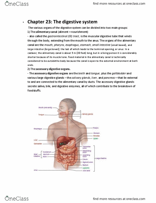
2. List the regions of the digestive system that food passes through
during digestion.
-Alimentary Canal:
oOral cavity to the anus
-Gastrointestinal tract:
oPart of the alimentary canal
oStomach & intestines (small and large)
-Accessory glands and structure:
oLiver
oPancreas
3. Identify the layers of the wall of the digestive tract
-Alimentary Canal Wall has FOUR layers:
oVariation occurs along the length of the GI tract
oFrom the esophagus to the anal canal, the wall has the same four layers
oMucosa: next to the lumen
Epithelium
Lamina propria
Muscularis mucosa
oSubmucosa
Fibrous connective tissue
Submucosal nerve plexus: controls gland secretions
oMuscularis
Two layers of muscle
Longitudinal muscle (goes in the same direction of the tube)
Circular muscle (goes around the tube)
oCauses lumen to get smaller
Myenteric Plexus: controls peristalsis
oSerosa: outer surface
Epithelium
find more resources at oneclass.com
find more resources at oneclass.com

MIDTERM 2 WEEK 7 OBJECTIVES
Connective tissue
-Peritoneum & Mesenteries:
oPeritoneum: serious membrane lining the abdominopelvic cavity and
covering the surfaces of the organs within
Visceral peritoneum: serosa
Parietal peritoneum: lines the cavity and is continuous with the
visceral peritoneum
Peritoneal cavity: space between containing serous fluid
Peritonitis: inflammation of the peritoneum
4. Name and describe the mesenteries
-Mesenteries: connective tissue sheets holding abdominal viscera in place
oDouble layers of serous membrane that will help to attach organs to the
dorsal wall of the abdomen
Functions
Provide routes for blood vessels, lymphatics, and nerves
Hold organs in place
Store fat, lymph nodes
oVery important for pathways of nerves and blood vessels to get to those
organs
-Lesser omentum: extends from lesser curvature of stomach to liver
oSuspends stomach and small intestine (duodenum portion) from liver
oConnects the stomach to the intestines
-Greater omentum: hangs down like an apron from stomach’s greater curvature
oDrapes over the ventral surface of the viscera
-Mesocolon: mesentery of the colon
oBinds large intestine to the posterior wall
find more resources at oneclass.com
find more resources at oneclass.com

MIDTERM 2 WEEK 7 OBJECTIVES
5. Describe the oral cavity and the structure of the teeth and
tongue
-Oral (Buccal) Cavity:
oIngestion
oDigestion
oTaste
oVoice-production
oSwallowing
oBreathing
-Lips and Cheeks: skeletal muscle (orbicularis oris) covered externally by skin
oBuccinators form cheeks
oInside lining is stratified squamous cell epithelium
oHelp keep food between teeth when we chew
oAid in speech
oLips are large
Extend from the inferior margin of the nose to the superior border of
the chin
What causes the “lips” to be red?
Lips don’t have a lot of keratin in it
Becomes more transparent, so blood vessels become more
apparent
-Palate: forms the roof of the mouth
oHard palate anterior
oSoft palate posterior
Uvula hangs down
Moves up when you swallow and blocks the nasopharynx so
food doesn’t go up your nasal cavity
Soft palate is anchored to the tongue
-Tongue: occupies the floor of the mouth; fills most of the oral cavity
oBundles of skeletal muscle = intrinsic muscles
oExtrinsic muscles: attach it to the skull and hyoid bone
oLingual frenulum: secures it to the floor
oMucosa covering
oPapillae (bumps on the tongue)
Filiform papillae
Fungiform papilla
find more resources at oneclass.com
find more resources at oneclass.com
Document Summary
Identify the functions and major processes of the digestive system. Ingestion: intake of food: consolidation of indigestible residue. Defecation: elimination of feces: list the regions of the digestive system that food passes through during digestion. Alimentary canal: oral cavity to the anus. Gastrointestinal tract: part of the alimentary canal, stomach & intestines (small and large) Accessory glands and structure: liver, pancreas. Identify the layers of the wall of the digestive tract. Alimentary canal wall has four layers: variation occurs along the length of the gi tract, from the esophagus to the anal canal, the wall has the same four layers, mucosa: next to the lumen. Submucosal nerve plexus: controls gland secretions: muscularis. Longitudinal muscle (goes in the same direction of the tube) Circular muscle (goes around the tube: causes lumen to get smaller. Myenteric plexus: controls peristalsis: serosa: outer surface. Peritoneum & mesenteries: peritoneum: serious membrane lining the abdominopelvic cavity and covering the surfaces of the organs within.