NURS 301 Lecture 6: Online Lecture - Week 6 (Blood)


Blood Functions
Transport
Transport material around the body and get to and away
from cells
○
Taking nutrients like glucose, salts, vitamins to cells
○
Taking away waste products from the cell
○
-
Homeostasis
pH
○
Tb (basal body temperature)
Within a normal temperature
§
○
Hydration of cells
Blood is primarily water
§
○
-
Protection against fluid loss (clotting) and disease (white blood
cells)
-
Blood Characteristics
Fluid connective tissue
-
pH range: 7.35-7.45 (slightly basic)
-
Volume: 5-6 liters in males, 4-5 liters in females
-
Plasma (liquid portion) + Formed Elements (Packed Cell
Volume)
-
Components and general properties of blood
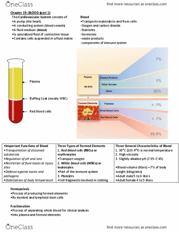
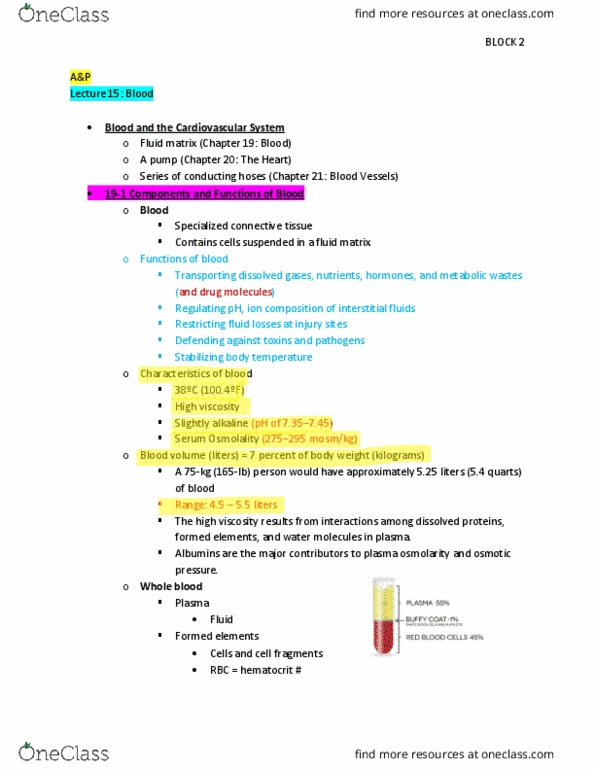
Plasma: clear, extracellular fluid, matrix
55% of whole blood
Water (92%)
□
Suspended substances
Plasma proteins
®
Ions
®
Nutrients, hormones, and waste products
®
Dissolved gases
®
□
§
○
Formed elements: cells and cell fragments
45% of whole blood
Erythrocytes (red blood cells)
□
Leukocytes (white blood cells)
□
Platelets (thrombocytes)
□
§
Myeloid hemopoiesis occurs in red bone
All types of formed elements produced in red
bone marrow
□
§
○
-
There are more erythrocytes
-
Types of Plasma Proteins
Albumin (60%)
Colloidal osmotic pressure
Filtration or flow of water
§
Greater pick up of fluid of tissues into capillaries
§
Liver disease
Reduce proteins in capillaries and plasma
□
Less fluid pick up
□
More fluid building up in interstitial fluid
Causes edema
®
□
§
○
Buffer
○
Transport (bilirubin)
Carry material around in the blood
§
Lipids and hormones are not soluble, must attach to
molecule to flow through
§
○
-
Globulins (36%)
Defense against invading bacteria
○
Immunoglobulins: antibodies for body defense against
infection
○
Compliment: chemicals for defense against infection
○
Transport
Transferrin: transport iron to liver for example for
storage
§
Lipoproteins (VLDL, LDL, IDL, HDL): carry
cholesterol around in body
§
○
Others
○
-
Fibrinogen (4%)
Blood clotting protein
○
-
Erythrocytes
Function to transport oxygen; secondarily to transport CO2
-
About 5 million per microliter of blood
-
Erythropoiesis in red bone marrow of long bones, ribs,
sternum after puberty, spleen and liver of fetus
-
Hormones stimulate erythropoiesis
Erythropoietin a peptide hormone from kidneys, released
due to hypoxia (low tissue oxygen level)
Hypoxia stimulates the release of erythropoietin
§
If you have a low level of oxygen, blood flowing to
the kidneys, there are sensory structures that pick up
the level of oxygens
When it is low, kidney releases more
erythropoietin and flows through blood stream,
reaches red bone, releases more red blood cells
□
§
○
Testosterone
○
-
No nucleus
-
Size: slight smaller than capillaries
-
Biconcave disc
Maximizes surface area to volume ratio
○
Decrease diffusion distance
○
Allows cell to flex when moving through capillaries
○
-
Flexible, thin
Easier for oxygen to get in when thin, easier diffusion
○
-
Erythrocyte Life Cycle
Erythropoiesis - production of RBC
Originate from hemopoietic stem cells
○
ECFU are stimulate by erythropoietin
Colony forming unit
§
○
-
Hemopoietic stem cell ---> CFU ---> erythroblast --->
reticulocyte ---> erythrocyte (mature cell)
-
Doing blood test with elevated reticulocytes
Anemia
○
-
Content of Erythrocyte
Lipids
-
ATP
-
Carbonic anhydrase (enzyme for buffering system)
-
Hemoglobin
4 different protein chains, snake like
With each one there is a iron-containing heme group
§
○
-
Hemoglobin Structure
Protein portion is Globin
Adult --- 4 polypeptide chains --- 2 alpha; 2 beta
○
Fetal hemoglobin contains --- 2 alpha; 2 gamma (greater
binding capacity compared to mom's)
○
-
Heme group (red pigment) contains iron
-
The structure of Hb allow it to reversibly bind 4 oxygen
molecules
For each heme group
○
-
Oxyhemoglobin (if it has oxygen)
eg. Picking up oxygen in lungs
○
-
deoxyhemoglobin (oxygen removed)
eg. Releasing oxygen to tissues
○
-
carbaminohemoglobin
(attach carbon dioxide as waste product)
○
-
Erythrocyte Recycling and Concentration in Blood
Because they lack a nucleus and many organelles, they are not
around for long
Parts of the cell will break down as they're used over time,
without nucleus there is no genetic material to build new
parts
○
120 days
○
Range 60-180 days
○
-
Concentration in blood
Females 4.2-5.4 million/microliter
○
Males 4.6-6.2 million/microliter
Testosterone stimulate erythropoiesis
§
○
-
Oxygen-Carrying Capacity of Blood
How many RBCs total in a male? Female?
Example: 5 million/microliter and 1million microliters/1
liter and 5 liters of blood in a person, 25 trillion blood
cells per person
○
-
There are about 270 million hemoglobin/erythrocyte
-
How much oxygen carried around?
Multiple 270 million time 25 trillion times four oxygen
per hemoglobin
○
-
Degrading Hemoglobin
Spleen, liver and red bone marrow
-
Macrophages
-
Globin ---> amino acids which are recycled into proteins
-
Heme ---> iron and biliverdin (green pigment)
Iron is carried by transferring to be stored in liver, bone,
spleen cells
○
Biliverdin ---> bilirubin ---> bilirubin/albumin ---> liver,
where it is used to make bile for digestion
○
-
Jaundice
Elevated levels of bilirubin lead to diffusion of excess into
peripheral tissues
-
Skin and whites of eyes yellow
-
Neonate jaundice
Newborns
○
Typical because of liver is immature, cannot function
quite yet
○
Premature infant, can also happen to full term
○
Can happen because of infection
○
-
Liver disease
Aren't able to process bilirubin into bile, spilling of
bilirubin into tissues
○
-
Treated with light therapy, breaks down excess bilirubin
-
Hematocrit: test for evaluation of packed cell volume, what % of
your whole blood, % of erythrocytes in blood
Normal values
Males 45%-52%
○
Females 37%-48%
○
-
Low is anemia
-
Elevate is polcythemia
-
Too few blood cells? ANEMIA
Oxygen carrying capacity of blood is reduced
-
Fatigue, cold-intolerance, pale
-
Causes: accelerated RBC loss or decreased production of RBCs
or hemoglobin (dietary deficiencies) or inadequate
erythropoietin production
-
Types of anemia due to genetic inheritance
Thalassemia: typically seen in people of Mediterranean
ancestry
Globin chains are absent or faulty leading to short-
lived RBCs
§
○
Sickle cell anemia: genetic disorder abnormal
hemoglobin distorts RBC shape
Instead of normal disc shape, there is a sickle shape
§
Can stick to inside of blood vessel, aggregate, cause
blocking of blood vessel
Heart attack, stroke
□
§
Found commonly in people of African descent
Linked to malaria
□
1 abnormal gene form, you don't have sickle
shape of cell
Malaria infection agent, less able to infect
RBC
®
□
Sickle cell trait is advantage for prevent malaria
□
§
○
-
Too many? POLYCYTHEMIA
Caused by cancer of red bone marrow
-
Causes blood to be more viscous, thicker
Harder for heart to move viscous blood through
circulatory system
○
Blockage of blood vessels
○
-
Treatment: donate blood
Remove level of RBCs
○
-
Blood doping
Athletes due to increase oxygen load in blood
○
More oxygen allows for more energy
○
Deliver blood, have some blood removed
Spin it down and have packed cell volume
§
Inject into blood stream
§
Increase overall RBC
§
○
Dangerous because it affects viscosity of blood
○
-
Blood Types
Several genetically determined blood groups with multiple
types
-
ABO and Rh most common
-
Red blood cell contains glycolipid antigen on membrane
Surface antigens made of carbohydrates and fat
○
Hanging off surface of cells
○
All have the same stem, difference is caused by chain
hanging on
○
Type O: no chain
○
-
Plasma contains antibodies that react against foreign antigens
-
Use antibodies against the different types
Antibodies are special proteins that cause reaction with
antigen and cause clumping
○
If you have the clumping if means you have the clumping
on your antigen (on your own)
○
-
Rh antigen is positive, don't have is negative
-
O has neither A or B antigens
Produced A and B antibodies to A and B blood
○
-
Type A has antigen A
Produce B type antibodies against B antigen
○
-
Type B has antigen B
Produce A type antibodies against A antigen
○
-
Type AB has antigen AB
Neither A nor B antibodies produced
○
-
Rh (D)
Another type of blood cell antigen
○
If you are Rh- and exposed to +
Then you produce Rh antibodies to attack them
§
○
Causes a situation during pregnancy
Potential dangerous
Rh- mom with Rh+ fetus (father Rh+)
□
Does not hurt first pregnancy
□
§
Material and fetal blood not mixing, separate
circulator system
When baby is delivered, hemorrhaging occurs,
blood from fetus is flowing into mother
□
§
Subsequent pregnancy, Mom produces antibodies
Next baby of exchanging tissues in placenta,
antibodies can attack baby's blood cells
□
§
○
-
Leukocytes
White blood cells
-
Fewer white blood cells than red blood cells
-
Contain nucleus and organelles
-
4000-11000/microliter of blood
-
General function is defense against foreign or abnormal
molecules and microorganisms that enter the body
-
Most are located in connective tissue proper or lymphatic
system. Only 2$ in the blood at any one time. They leave the
blood stream by squeezing between cells of capillary walls.
-
Movement due to chemotaxis
Cells damage release chemical that attracts them to area
○
-
Two major groups based on differential staining with Wright's
Stain
Granulocytes (granules stain dark)
Neutrophils, most common type in blood
§
Eosinophils, next most common
§
Basophils, very low
§
○
Agranulocytes (granules do not stain well)
Lymphocytes, second most prevalent after
neutrophils
§
monocytes
§
○
They store different chemicals in granules in use for
defense
○
-
Leukopoiesis
Production of white blood cells in red bone marrow
-
Hemopoietic
-
Most cells made in bone marrow, developed there, stored there,
until released
-
T lymphocytes mature in thymus
-
Leukocyte life span ranges: some live only days, others live for
decades
Involved in defense against invaders, viruses, and bacteria
○
-
Platelet Production
Thrombopoiesis
Some hematopoietic stem cells become megakaryoblasts
○
Megakaryocytes sprout proplatelet tendrils in red marrow
○
Many proplatelets are broken into platelets within lung
capillaries
○
-
Hemostasis
Cessation of bleeding, stop bleeding
-
Platelets release serotonin and clotting factors
-
Three stage
Serotonin triggers vasoconstriction
○
Platelet plug seals vessel
○
Clotting factors convert fibrinogen to sticky fibrin
○
-
Once crisis passes, platelets secrete growth factors to trigger
healing and other factors that cause dissolving of the clot
-
Phase 1: vascular phase, vascular spasm
-
Phase 2: platelet phase, platelet aggregation
Stimulating pathway for clot production
○
-
Clot blocks off tissue so bacteria cannot infiltrate further into
tissue
-
Bacteria can product chemical streptokinase digest fibrin
allowing bacteria to pass through the clot
-
Online Lecture - Week 6 (Blood)
Sunday, April 29, 2018
7:44 PM

Blood Functions
Transport
Transport material around the body and get to and away
from cells
○
Taking nutrients like glucose, salts, vitamins to cells
○
Taking away waste products from the cell
○
-
Homeostasis
pH
○
Tb (basal body temperature)
Within a normal temperature
§
○
Hydration of cells
Blood is primarily water
§
○
-
Protection against fluid loss (clotting) and disease (white blood
cells)
-
Blood Characteristics
Fluid connective tissue
-
pH range: 7.35-7.45 (slightly basic)
-
Volume: 5-6 liters in males, 4-5 liters in females
-
Plasma (liquid portion) + Formed Elements (Packed Cell
Volume)
-
Components and general properties of blood
Plasma: clear, extracellular fluid, matrix
55% of whole blood
Water (92%)
□
Suspended substances
Plasma proteins
®
Ions
®
Nutrients, hormones, and waste products
®
Dissolved gases
®
□
§
○
Formed elements: cells and cell fragments
45% of whole blood
Erythrocytes (red blood cells)
□
Leukocytes (white blood cells)
□
Platelets (thrombocytes)
□
§
Myeloid hemopoiesis occurs in red bone
All types of formed elements produced in red
bone marrow
□
§
○
-
There are more erythrocytes
-
Types of Plasma Proteins
Albumin (60%)
Colloidal osmotic pressure
Filtration or flow of water
§
Greater pick up of fluid of tissues into capillaries
§
Liver disease
Reduce proteins in capillaries and plasma
□
Less fluid pick up
□
More fluid building up in interstitial fluid
Causes edema
®
□
§
○
Buffer
○
Transport (bilirubin)
Carry material around in the blood
§
Lipids and hormones are not soluble, must attach to
molecule to flow through
§
○
-
Globulins (36%)
Defense against invading bacteria
○
Immunoglobulins: antibodies for body defense against
infection
○
Compliment: chemicals for defense against infection
○
Transport
Transferrin: transport iron to liver for example for
storage
§
Lipoproteins (VLDL, LDL, IDL, HDL): carry
cholesterol around in body
§
○
Others
○
-
Fibrinogen (4%)
Blood clotting protein
○
-
Erythrocytes
Function to transport oxygen; secondarily to transport CO2
-
About 5 million per microliter of blood
-
Erythropoiesis in red bone marrow of long bones, ribs,
sternum after puberty, spleen and liver of fetus
-
Hormones stimulate erythropoiesis
Erythropoietin a peptide hormone from kidneys, released
due to hypoxia (low tissue oxygen level)
Hypoxia stimulates the release of erythropoietin
§
If you have a low level of oxygen, blood flowing to
the kidneys, there are sensory structures that pick up
the level of oxygens
When it is low, kidney releases more
erythropoietin and flows through blood stream,
reaches red bone, releases more red blood cells
□
§
○
Testosterone
○
-
No nucleus
-
Size: slight smaller than capillaries
-
Biconcave disc
Maximizes surface area to volume ratio
○
Decrease diffusion distance
○
Allows cell to flex when moving through capillaries
○
-
Flexible, thin
Easier for oxygen to get in when thin, easier diffusion
○
-
Erythrocyte Life Cycle
Erythropoiesis - production of RBC
Originate from hemopoietic stem cells
○
ECFU are stimulate by erythropoietin
Colony forming unit
§
○
-
Hemopoietic stem cell ---> CFU ---> erythroblast --->
reticulocyte ---> erythrocyte (mature cell)
-
Doing blood test with elevated reticulocytes
Anemia
○
-
Content of Erythrocyte
Lipids
-
ATP
-
Carbonic anhydrase (enzyme for buffering system)
-
Hemoglobin
4 different protein chains, snake like
With each one there is a iron-containing heme group
§
○
-
Hemoglobin Structure
Protein portion is Globin
Adult --- 4 polypeptide chains --- 2 alpha; 2 beta
○
Fetal hemoglobin contains --- 2 alpha; 2 gamma (greater
binding capacity compared to mom's)
○
-
Heme group (red pigment) contains iron
-
The structure of Hb allow it to reversibly bind 4 oxygen
molecules
For each heme group
○
-
Oxyhemoglobin (if it has oxygen)
eg. Picking up oxygen in lungs
○
-
deoxyhemoglobin (oxygen removed)
eg. Releasing oxygen to tissues
○
-
carbaminohemoglobin
(attach carbon dioxide as waste product)
○
-
Erythrocyte Recycling and Concentration in Blood
Because they lack a nucleus and many organelles, they are not
around for long
Parts of the cell will break down as they're used over time,
without nucleus there is no genetic material to build new
parts
○
120 days
○
Range 60-180 days
○
-
Concentration in blood
Females 4.2-5.4 million/microliter
○
Males 4.6-6.2 million/microliter
Testosterone stimulate erythropoiesis
§
○
-
Oxygen-Carrying Capacity of Blood
How many RBCs total in a male? Female?
Example: 5 million/microliter and 1million microliters/1
liter and 5 liters of blood in a person, 25 trillion blood
cells per person
○
-
There are about 270 million hemoglobin/erythrocyte
-
How much oxygen carried around?
Multiple 270 million time 25 trillion times four oxygen
per hemoglobin
○
-
Degrading Hemoglobin
Spleen, liver and red bone marrow
-
Macrophages
-
Globin ---> amino acids which are recycled into proteins
-
Heme ---> iron and biliverdin (green pigment)
Iron is carried by transferring to be stored in liver, bone,
spleen cells
○
Biliverdin ---> bilirubin ---> bilirubin/albumin ---> liver,
where it is used to make bile for digestion
○
-
Jaundice
Elevated levels of bilirubin lead to diffusion of excess into
peripheral tissues
-
Skin and whites of eyes yellow
-
Neonate jaundice
Newborns
○
Typical because of liver is immature, cannot function
quite yet
○
Premature infant, can also happen to full term
○
Can happen because of infection
○
-
Liver disease
Aren't able to process bilirubin into bile, spilling of
bilirubin into tissues
○
-
Treated with light therapy, breaks down excess bilirubin
-
Hematocrit: test for evaluation of packed cell volume, what % of
your whole blood, % of erythrocytes in blood
Normal values
Males 45%-52%
○
Females 37%-48%
○
-
Low is anemia
-
Elevate is polcythemia
-
Too few blood cells? ANEMIA
Oxygen carrying capacity of blood is reduced
-
Fatigue, cold-intolerance, pale
-
Causes: accelerated RBC loss or decreased production of RBCs
or hemoglobin (dietary deficiencies) or inadequate
erythropoietin production
-
Types of anemia due to genetic inheritance
Thalassemia: typically seen in people of Mediterranean
ancestry
Globin chains are absent or faulty leading to short-
lived RBCs
§
○
Sickle cell anemia: genetic disorder abnormal
hemoglobin distorts RBC shape
Instead of normal disc shape, there is a sickle shape
§
Can stick to inside of blood vessel, aggregate, cause
blocking of blood vessel
Heart attack, stroke
□
§
Found commonly in people of African descent
Linked to malaria
□
1 abnormal gene form, you don't have sickle
shape of cell
Malaria infection agent, less able to infect
RBC
®
□
Sickle cell trait is advantage for prevent malaria
□
§
○
-
Too many? POLYCYTHEMIA
Caused by cancer of red bone marrow
-
Causes blood to be more viscous, thicker
Harder for heart to move viscous blood through
circulatory system
○
Blockage of blood vessels
○
-
Treatment: donate blood
Remove level of RBCs
○
-
Blood doping
Athletes due to increase oxygen load in blood
○
More oxygen allows for more energy
○
Deliver blood, have some blood removed
Spin it down and have packed cell volume
§
Inject into blood stream
§
Increase overall RBC
§
○
Dangerous because it affects viscosity of blood
○
-
Blood Types
Several genetically determined blood groups with multiple
types
-
ABO and Rh most common
-
Red blood cell contains glycolipid antigen on membrane
Surface antigens made of carbohydrates and fat
○
Hanging off surface of cells
○
All have the same stem, difference is caused by chain
hanging on
○
Type O: no chain
○
-
Plasma contains antibodies that react against foreign antigens
-
Use antibodies against the different types
Antibodies are special proteins that cause reaction with
antigen and cause clumping
○
If you have the clumping if means you have the clumping
on your antigen (on your own)
○
-
Rh antigen is positive, don't have is negative
-
O has neither A or B antigens
Produced A and B antibodies to A and B blood
○
-
Type A has antigen A
Produce B type antibodies against B antigen
○
-
Type B has antigen B
Produce A type antibodies against A antigen
○
-
Type AB has antigen AB
Neither A nor B antibodies produced
○
-
Rh (D)
Another type of blood cell antigen
○
If you are Rh- and exposed to +
Then you produce Rh antibodies to attack them
§
○
Causes a situation during pregnancy
Potential dangerous
Rh- mom with Rh+ fetus (father Rh+)
□
Does not hurt first pregnancy
□
§
Material and fetal blood not mixing, separate
circulator system
When baby is delivered, hemorrhaging occurs,
blood from fetus is flowing into mother
□
§
Subsequent pregnancy, Mom produces antibodies
Next baby of exchanging tissues in placenta,
antibodies can attack baby's blood cells
□
§
○
-
Leukocytes
White blood cells
-
Fewer white blood cells than red blood cells
-
Contain nucleus and organelles
-
4000-11000/microliter of blood
-
General function is defense against foreign or abnormal
molecules and microorganisms that enter the body
-
Most are located in connective tissue proper or lymphatic
system. Only 2$ in the blood at any one time. They leave the
blood stream by squeezing between cells of capillary walls.
-
Movement due to chemotaxis
Cells damage release chemical that attracts them to area
○
-
Two major groups based on differential staining with Wright's
Stain
Granulocytes (granules stain dark)
Neutrophils, most common type in blood
§
Eosinophils, next most common
§
Basophils, very low
§
○
Agranulocytes (granules do not stain well)
Lymphocytes, second most prevalent after
neutrophils
§
monocytes
§
○
They store different chemicals in granules in use for
defense
○
-
Leukopoiesis
Production of white blood cells in red bone marrow
-
Hemopoietic
-
Most cells made in bone marrow, developed there, stored there,
until released
-
T lymphocytes mature in thymus
-
Leukocyte life span ranges: some live only days, others live for
decades
Involved in defense against invaders, viruses, and bacteria
○
-
Platelet Production
Thrombopoiesis
Some hematopoietic stem cells become megakaryoblasts
○
Megakaryocytes sprout proplatelet tendrils in red marrow
○
Many proplatelets are broken into platelets within lung
capillaries
○
-
Hemostasis
Cessation of bleeding, stop bleeding
-
Platelets release serotonin and clotting factors
-
Three stage
Serotonin triggers vasoconstriction
○
Platelet plug seals vessel
○
Clotting factors convert fibrinogen to sticky fibrin
○
-
Once crisis passes, platelets secrete growth factors to trigger
healing and other factors that cause dissolving of the clot
-
Phase 1: vascular phase, vascular spasm
-
Phase 2: platelet phase, platelet aggregation
Stimulating pathway for clot production
○
-
Clot blocks off tissue so bacteria cannot infiltrate further into
tissue
-
Bacteria can product chemical streptokinase digest fibrin
allowing bacteria to pass through the clot
-
Online Lecture - Week 6 (Blood)
Sunday, April 29, 2018 7:44 PM
Document Summary
Transport material around the body and get to and away from cells. Taking nutrients like glucose, salts, vitamins to cells. Protection against fluid loss (clotting) and disease (white blood cells) Fluid connective tissue ph range: 7. 35-7. 45 (slightly basic) Volume: 5-6 liters in males, 4-5 liters in females. Plasma (liquid portion) + formed elements (packed cell. All types of formed elements produced in red bone marrow. Greater pick up of fluid of tissues into capillaries. Lipids and hormones are not soluble, must attach to molecule to flow through. Transferrin: transport iron to liver for example for storage. Lipoproteins (vldl, ldl, idl, hdl): carry cholesterol around in body. Function to transport oxygen; secondarily to transport co2. Erythropoiesis in red bone marrow of long bones, ribs, sternum after puberty, spleen and liver of fetus. Erythropoietin a peptide hormone from kidneys, released due to hypoxia (low tissue oxygen level)