PHTY100 Lecture Notes - Lecture 7: Abductor Digiti Minimi Muscle Of Hand, Extensor Retinaculum Of The Hand, Achilles Tendon


Muscles of the Leg and Foot
7.1 Identify, list attachments and deduce the action(s) of the muscles of the leg:
•Anterior group
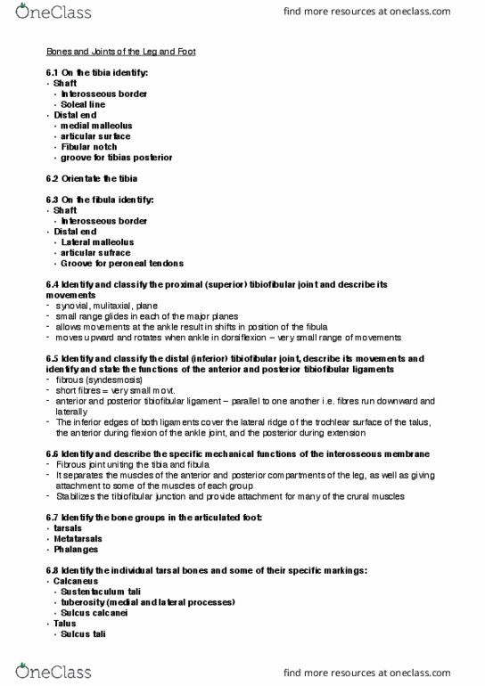
•Tibialis anterior
•PA: lateral proximal 2/3 of tibia, adjacent interosseous membrane
•DA: plantar surface of the medial cuneiform, base of metatarsal 1
•A: dorsiflexes foot at ankle, inverts foot
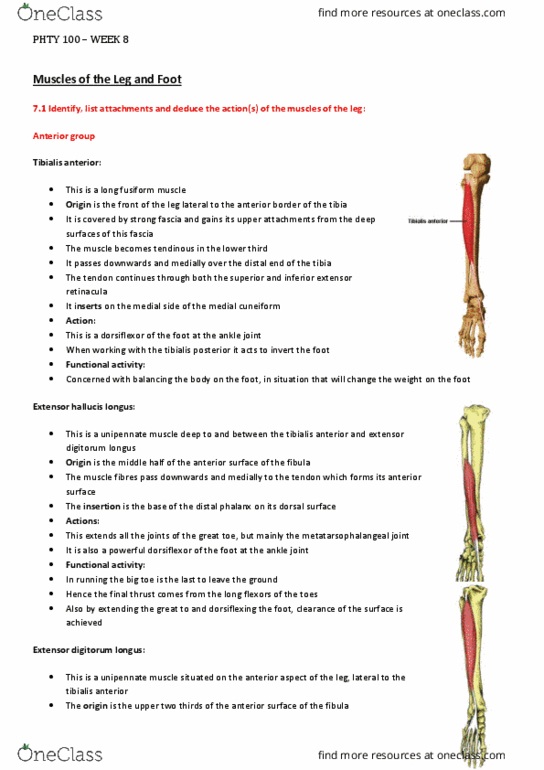
•Extensor hallucis longus
•PA: middle 1/2 anterior fibula, adjacent interosseous membrane
•DA: dorsal surface of base of distal phalanx of digit 1
•A: dorsiflexes foot at ankle, extends 1st interphalangeal joint
•Extensor digitorum longus
•PA: proximal 2/3 anterior fibula, adjacent interosseous membrane, lateral condyle of tibia
•DA: bases of middle and distal phalanges of digits 2-5 via dorsal digital expansion (extensor
aponeurosis/hood)
•A: Extends 2nd-5th distal interphalangeal joints
•Peroneus Tertius
•PA: anterior distal 1/4 of fibula, adjacent interosseus membrane
•DA: dorsal surface of base of metatarsal 5
•A: Dorsiflexes foot at ankle, everts foot
•Lateral group
•Peroneus longus
•PA: lateral surface proximal 2/3 and head of fibula
•DA: medial cuneiform, base of metatarsal 1
•A: plantarflexes foot at ankle, everts foot
•Peroneus brevis
•PA: lateral surface distal 2/3 of fibula
•DA: tuberosity on base of metatarsal 5
•A: plantarflexes foot at ankle, everts foot
•Posterior group
•gastrocnemius
•PA: posterior aspect of the medial and lateral femoral condyles, capsule of knee joint
•DA: posterior calcaneus via tendocalcaneus
•A: Plantar flexes foot at ankle
•Soleus
•PA: soleal line and posterior surface of The tibia, posterior surface upper third of fibula
•DA: posterior calcaneus via tendocalcaneus
•A: plantarflexes foot
•Plantaris
•PA: lateral supracondylar ridge of the femur, knee joint capsule
•DA: posterior calcaneus
•A: plantarflexes foot
•Popliteus
•PA: lateral condyle of the femur, lateral meniscus of the knee
•DA: posterior tibia above the soleal line
•A: flexes lower leg, medially rotates lower leg
•Tibialis posterior
•PA: proximal half of the posterior tibia below the soleal line, proximal two thirds of the fibula,
adjacent interosseus membrane
•DA: tubercle of the navicular, tendinous expansions connect to all tarsal bones (except the
talus) and the bases of the middle three metatarsals
•A: inverts foot
•Flexor digitorum longus
•PA: proximal half of the medial tibia below the soleal line
•DA: plantar surface of the base of the distal phalanx of digits 2 - 5
find more resources at oneclass.com
find more resources at oneclass.com