PHTY101 Lecture Notes - Lecture 3: Aponeurosis, Triangular Fibrocartilage, Axial Skeleton


capitulum
trochlear
medial and lateral epicondyles
medial and lateral supracondylar ridges
radial, ulnar and olecranon fossae
distal end
○
3.1 On the humerus, (re)identify:
•
head
neck
radial tuberosity
proximal end
○
impression for pronator teres
shaft
○
ulnar notch
distal end
○
3.2 Identify and classify the radius, and identify:
•
olecranon process
coronoid process
trochlear notch
radial notch
ulnar tuberosity
supinator crest
proximal end
○
shaft
○
head
styloid process
distal end
○
3.3 Identify and classify the ulna, and identify:
•
Classification: synovial, uniaxial, hinge
Flexion/extension (transverse axis)
□
Movements
elbow joint
○
Classification: synovial, uniaxial, pivot
□
Pronation/supination (longitudinal axis)
Movements
□
proximal (superior) radioulnar joint
Classification: synovial, uniaxial, pivot
□
Pronation/supination (longitudinal axis)
Movements
□
distal (inferior) radioulnar joint
radioulnar articulations
○
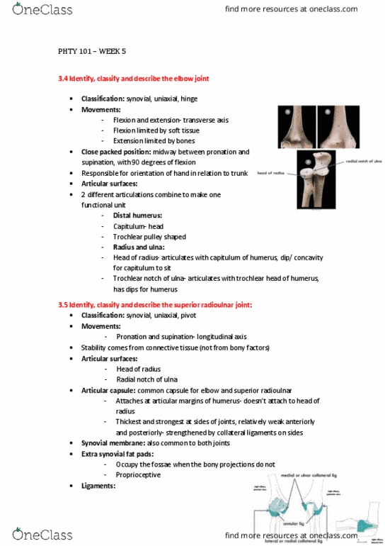
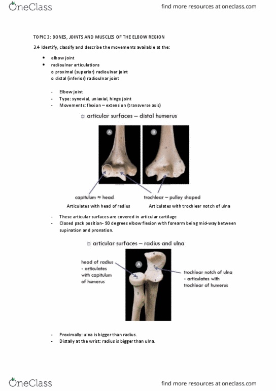
3.4 Identify, classify and describe the movements available at the:
•
Flexed 90 degrees
Forearm midway between supination and pronation (neutral)
CPP
○
Capitulum - anterior surface of humerus
□
Starts anteriorly, goes distally and goes posteriorly
Trochlear
□
Distal humerus
articular surfaces
○
3.5 At the elbow joint identify and/or describe its:
•
3. Bones, joints and muscles of the elbow region
Tuesday, 1 August 2017
2:14 PM
ANAT102 Page 1
find more resources at oneclass.com
find more resources at oneclass.com

Small concavity where capitulum sits
Rim is also articular with proximal radial joint
Head of radius
□
Two articular surfaces
Rougheed it i iddle whih does’t artiulate
Trochlear notch
□
Radius and ulna
Radius and ulna
articular surfaces
○
3.6 At the superior radioulnar joint identify and/or describe its:
•
Triangular in shape
Capsular
Collateral ligaments tense throughout flexion/extension range
○
3.5 and 3.6. ligaments
•
ANAT102 Page 2
find more resources at oneclass.com
find more resources at oneclass.com

Capsular
Prevents abduction
□
Medial epicondyle to back and front of joint
□
ulnar (medial) collateral
Prevents adduction
□
Lateral epicondyle to annular ligament
□
radial (lateral) collateral
Ring
Head of radius sits within ring - forms part of the articular surface
Anterior margin of the notch back to the posterior margin of the notch on the
ulnar
Upper part of ligament fuses to the capsule
Surrounds the head of the radius
Allows pronation/supination
Wider superiorly than inferiorly, funnel shaped
Prevents inferior dislocation of the head of the radius
Annular (RU joint)
○
Attaches at articular margins of humerus
Attaches to articular margins of ulna
No direct attachment to radius
Thickest and strongest at sides of joints
Relatively weak anteriorly, weak posteriorly
Capsule
○
Common to both joints
Tucks in under with an extra fold to allow pivoting of head of radius
Synovial membrane
○
Occupy the fossa when the bony projections don't
Flexion and extension pushes fat pads up and out
Proprioceptive nerve endings
Extra synovial fat pads
○
3.7 Describe the structural relationship between the elbow joint and the superior radioulnar
joint
•
3.8 At the inferior radioulnar joint identify and/or describe its:
•
ANAT102 Page 3
find more resources at oneclass.com
find more resources at oneclass.com
Document Summary
Bones, joints and muscles of the elbow region. 3. 1 on the humerus, (re)identify: distal end capitulum trochlear medial and lateral epicondyles medial and lateral supracondylar ridges radial, ulnar and olecranon fossae. 3. 2 identify and classify the radius, and identify: proximal end shaft head neck radial tuberosity impression for pronator teres distal end ulnar notch. 3. 3 identify and classify the ulna, and identify: proximal end olecranon process coronoid process trochlear notch radial notch ulnar tuberosity supinator crest shaft distal end head styloid process. 3. 4 identify, classify and describe the movements available at the: elbow joint. Flexion/extension (transverse axis) radioulnar articulations proximal (superior) radioulnar joint. 3. 5 at the elbow joint identify and/or describe its: Forearm midway between supination and pronation (neutral) articular surfaces. Rim is also articular with proximal radial joint. 3. 6 at the superior radioulnar joint identify and/or describe its: articular surfaces. Medial epicondyle to back and front of joint radial (lateral) collateral.