ANAT30008 Lecture Notes - Lecture 21: Anterior Cranial Fossa, Middle Meningeal Artery, Middle Cranial Fossa

16 Oct 2018
School
Department
Course
Professor

Document Summary
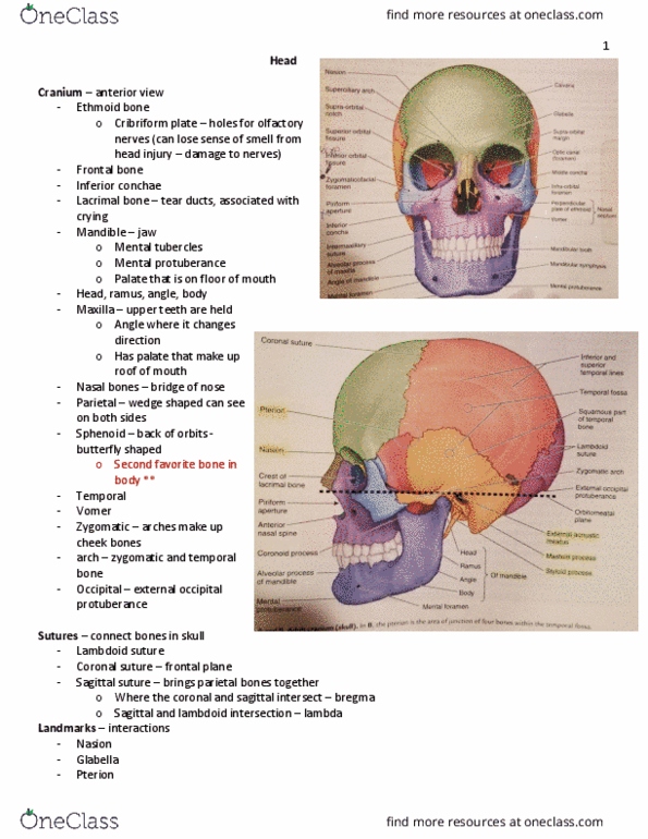
Growth of mandible and facial skeleton: in infants, cranium to face ratio is large; many of the bones haven"t yet fused, fontanelles = cartilaginous plate; anterior and posterior. Anterior clinically not palpable by 18 months, posterior not palpable by end of rst year: mandible fuses early in 2nd year. Sutures (4: sagittal, coronal, lambdoid and pterion, pterion = sutures from spenoid, parietal, temporal and frontal bones meet, quite weak if have lateral blow to head fracture and damage blood vessel that lies underneath haemorrhage. Cranial cavity: contains the brain and has three parts (anterior, posterior and middle fossae, also has openings for cranial nerves (and other things e. g. vessels) Anterior cranial fossa: has crista gali with cribriform plate of ethmoid bone (foramina open into nasal cavity to allow passage of sensory nerves from nose) olfactory nerves. Note: all nerves for eye other than optic run through cavernous sinus. Come o ventral surface of brain stem essentially in order.