BIOM20001 Lecture Notes - Lecture 5: Protein Folding, Supersecondary Structure, Beta Sheet

20 Jul 2018
School
Department
Course
Professor
Document Summary
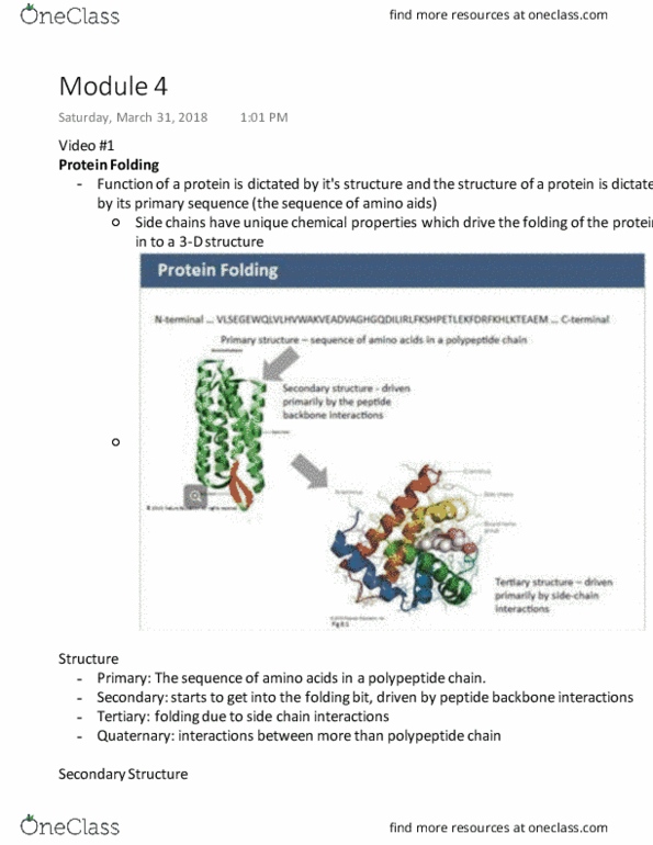
Draw diagrams illustrating the hydrogen bonding pattern in alpha-helices and in beta sheets. To turn alpha (alpha hairpins) and beta sheets. Consists of 4 residues (i, i+1, i+2, i+3) a. b. Draw diagrams illustrating the differences between parallel and antiparallel beta sheets. Using diagrams where appropriate explain the difference between primary, secondary, tertiary and quaternary protein structure. Define domain in terms of structure and evolution a. Independent folding, independent hydrophobic core, specific functions to each domains can be attributed. What genetic processes create proteins with different functional properties using existing domain structures a. What is meant by the statement that protein sequences can be optimally aligned a. If sequences of protein domains show >25% sequence identity, what can you say about their ancestry and structural similarity? a. Any polar group buried in the protein must form a hydrogen bond. This will include charged groups, but these are rarely buried.