BIOM20002 Lecture Notes - Lecture 40: Jejunum, Curvatures Of The Stomach, Abdominal Wall

Document Summary
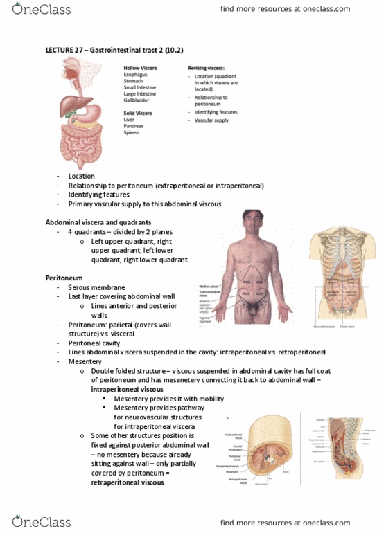
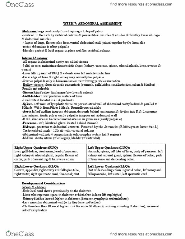
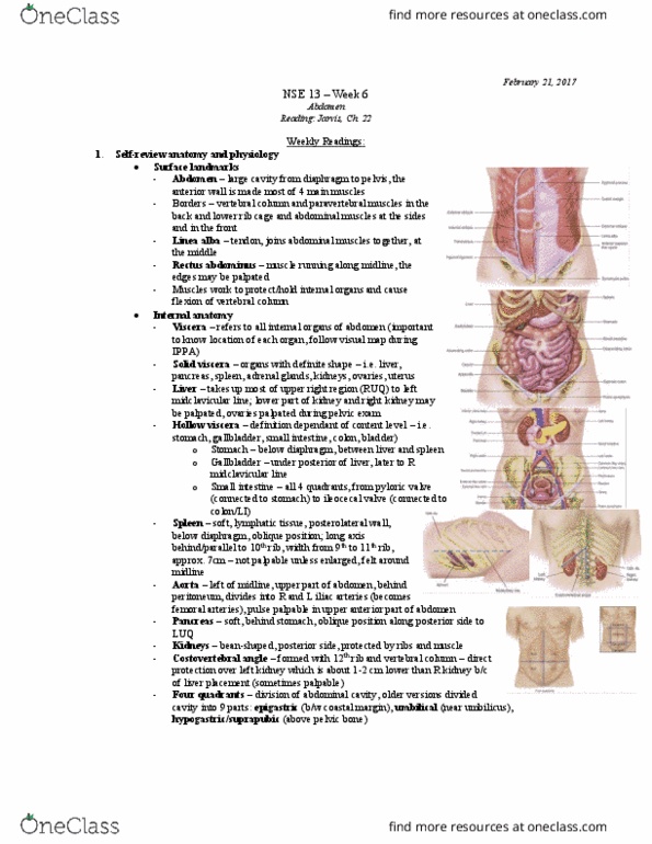
Horizontal plane and sagittal plane divides abdominal cavity into 4 quadrants. Peritoneal cavity: filled with viscera, contains serous fluid. Intraperitoneal viscus (ie. jejunum): suspended by mesentery, mobile. Retroperitoneal viscus: (ie. kidney): fixed, situated against abdominal wall. Only the peritoneal surface is covered by visceral peritoneum. Starts from c6, passes through diaphragm and enters stomach from right hand side. A muscular tube with narrowings at beginning, end and middle. Thoracic constriction: (at t4, t5) 1) pressure from bifurcation of trachea. 2) aortic arch goes up/back/left, produces impression on the left of esophagus - t4/5. Diaphragmatic constriction t10: passes through muscular part of diaphragm - a functional sphincter. Muscular tube covered by 2 complete layers of muscle. Z-line indicates where esophageal mucosa changes distinctly to stomach mucosa. Two orifices: 1) from esophagus (cardiac opening), 2) to duodenum (pyloric opening) Two curvatures: lesser curvature on top, greater curvature on bottom - j shaped morphology.