PHYS20008 Lecture Notes - Lecture 16: Heart Valve, Tricuspid Valve, Pulmonary Vein

12 Jun 2018
School
Department
Course
Professor

Lecture 16
PHYS20008 - HUMAN PHYSIOLOGY
LECTURE 16
THE CARDIAC CYCLE
OVERVIEW
•Relate structure to function
•Brief overview
•Conduction of message
•Path taken
•ECG
•Pressure changes
•Sequence of events
•Function of valves
•Combine it all together.
STRUCTURE & FUNCTION OF CARDIAC
CYCLE
•How is pumping co-ordinated and efficiency
maximized?
•Regular heart beat?
•Atria and ventricles?
•Blood squeezed out effectively?
•Back-flow prevented?
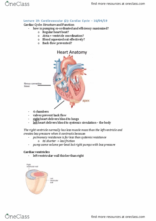
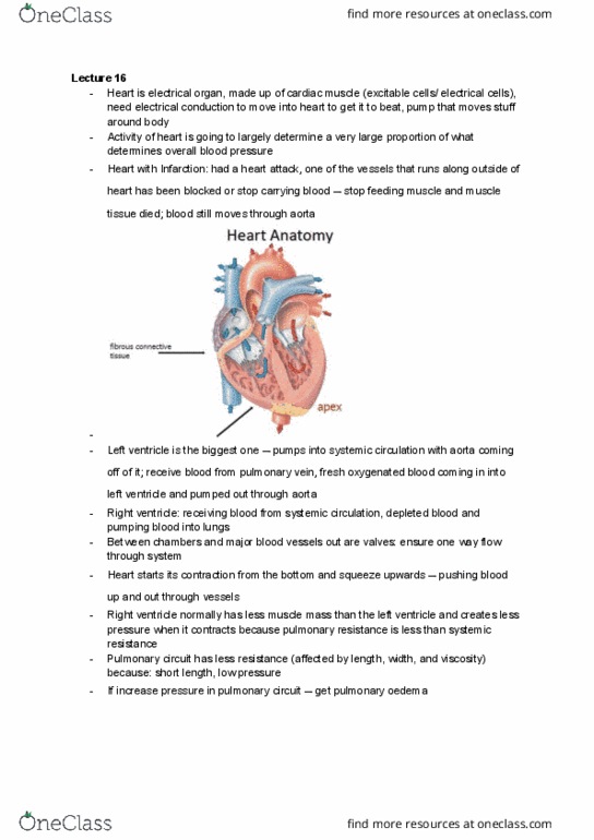
HEART ANATOMY
•Left ventricle is big one because it is
pumping into systemic circulation, with the
aorta coming off it. It receives blood from
the pulmonary vein.
•Right ventricle receives depleted blood
from systemic circulation and pumps it
into the lungs.
•We have valves ensuring we have one way
flow throughout the system.
•Q. Differences between right and left
ventricles:
•A. Pulmonary resistance is less than
systemic resistance
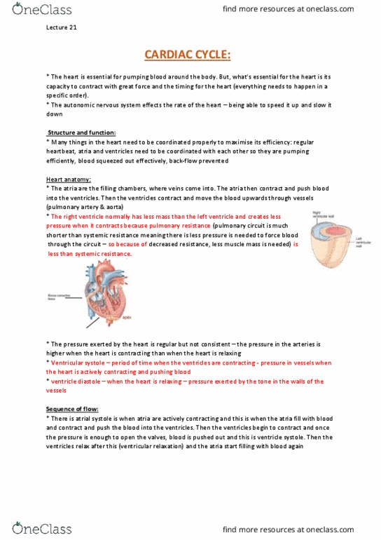
CARDIAC VENTRICLES
•Wall thickness?
•Force generated?
•Ventricular systole is contraction of the ventricles
•Ventricular diastole is relaxation of the ventricles
•Forcing blood through system in pulsating manner.
FROM BIOL10004
•The mammalian heart consists of four separate chambers, that alternately contract (systole) and
relax (diastole). Blood from the body enters the right atrium, moves through the tricuspid valve
into the right ventricle, and then through the pulmonary semilunar valve to the lung.

Lecture 16
PHYS20008 - HUMAN PHYSIOLOGY
•At the same time, blood returning from the lung enters the left atrium, goes through the bicuspid/
mitral valve into the left ventricle and then out through the aortic semilunar valve into the aorta.
•Valves control the direction of blood flow; they stop it going in the wrong direction.
•The tricuspid and mitral valves are strengthened by fibrous attachments, chordae tendinae, to the
ventricular wall, but the semilunar valves are not.
•The closing of the two valves is what causes the familiar heart sound; ‘lubb-dupp’. ‘lubb’ is due
to simultaneous closure of the tricuspid and mitral valves when the ventricles begin to contract.
The semilunar valves then open silently and blood is forced into the two major arteries. At the end
of the ventricular systole (contraction), pressure in the arteries causes the semilunar valves to close
abruptly, causing the ‘dupp’ sound.
•In humans, atrial contraction does not contribute greatly to the filling of the ventricles, because
the ventricles largely fill themselves by creating suction during diastole (relaxation), when the
relaxing ventricular walls passively expand, and also because there are no valves to prevent blood
from passing back into the veins. (However fish do have this)
CONDUCTION SYSTEM OF THE HEART
•Sinoatrial (SA) node is the pacemaker.
•Autorhythmic
•Pacemaker ~70 bpm
•Other areas
•Atrioventricular (AV) node (50 bpm)
•Purkinje fibers (25-40 bpm)
•
•The SA is at a higher rate, is the chief, running the show.
Document Summary
Overview: relate structure to function, brief overview, conduction of message, path taken, ecg, pressure changes, sequence of events, function of valves, combine it all together. Heart anatomy: left ventricle is big one because it is pumping into systemic circulation, with the aorta coming off it. It receives blood from the pulmonary vein: right ventricle receives depleted blood from systemic circulation and pumps it into the lungs, we have valves ensuring we have one way flow throughout the system, q. Cardiac ventricles: wall thickness, force generated, ventricular systole is contraction of the ventricles, ventricular diastole is relaxation of the ventricles, forcing blood through system in pulsating manner. From biol10004: the mammalian heart consists of four separate chambers, that alternately contract (systole) and relax (diastole). Blood from the body enters the right atrium, moves through the tricuspid valve into the right ventricle, and then through the pulmonary semilunar valve to the lung.