PSYC10003 Lecture Notes - Lecture 7: Electromagnetic Spectrum, Color Blindness, Ganglion Cell


7. The Human Visual System
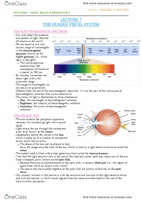
The Electromagnetic spectrum
• Eyes detect presence and pattern of light reflected off objects
• Sensitive to narrow range of wavelengths in the electromagnetic spectrum known as the visible
spectrum
o Extends from 380 nanometres to 760 nm
• Honeybees can detect light within UV range
• The range of wavelengths we can see is not qualitatively different from the rest of the
electromagnetic spectrum; just a part of the continuum of electromagnetic radiation we are sensitive
to
• Colour of light determined by 3 dimensions
1. Hue – wavelength of electromagnetic radiation
2. Brightness – intensity of electromagnetic radiation
3. Saturation – purity of electromagnetic radiation
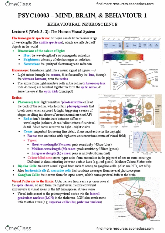
The Human Eye
• Eye contains peripheral
apparatus necessary for
transducing (transferring)
light into a neutral signal
Enters cornea:
transparent outer
layer
Lens: immediately
behind the cornea
and made up of a
number of
transparent layers
▪ Can be altered to help focus image onto the back of the eye which is lined by retina:
a light sensitive structure
Eyeball filled with vitreous humour: a clear gelatinous fluid
Light sensitive cells in retina (rods and cones) send their axons out of the eye from a common
point: optic disk
No photoreceptors in optic disk, causes blind spot (region of space from which an object is
not visible)
Axons that are bundled together at the optic disk are knock collectively as the optic nerve
find more resources at oneclass.com
find more resources at oneclass.com

2
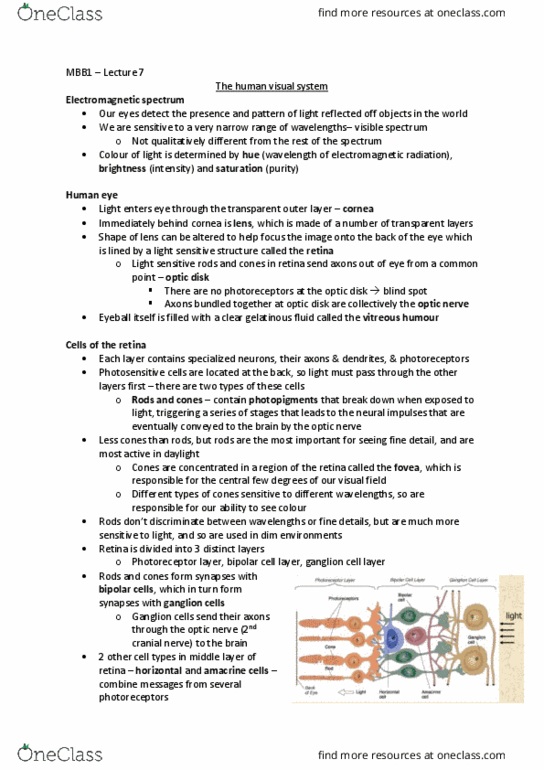
Cells of the retina
• close cross-section through the light sensitive retina reveals a series of layers
o each contain specialized neurons, their axons and dendrites, and the photoreceptors (the
retina is in fact a part of the brain)
• photosensitive (light sensitive) cells are located at the back of retina
o ∴ light must pass through each layer to get to them
• 2 types of photoreceptors:
1. rods: ≈120 million, dot disriiate between different wavelengths, cannot
discriminate fine visual detail, more sensitive to light than cones, so used in dimly lighted
environments
2. cones: ≈60 million, important for fine details, most active in daylight, concentrated in
fovea: responsible for the central degrees of our visual field, different types of cones
sensitive to different wavelengths of light, responsible for ability to see colour
• contain photopigments
o break down when exposed to light and this process triggers series of stages that lead to neural
impulses that are eventually conveyed to brain via optic nerve
• retina can be divided in 3 layers
1. Photoreceptor layer
2. Bipolar cell layer
3. Ganglion cell layer
rods and cones form
synapses with bipolar cells
which in turn form synapses
with ganglion cells
Ganglion cells send their
axons through optic nerve
(the second cranial nerve)
Conveying visual information
to brain
• Two other cells in the middle layer of retina: horizontal cells and amacrine cells, serve function of
combining messages from several photoreceptors
• Photoreceptors and bipolar cells do not produce AP but release neurotransmitters that increase or
decreases firing rate of AP generated by ganglion cells
Three Cone Types
1. Short-wavelength (S) cones –
peak sensitivity at 440 nm (blue light)
2. Medium-wavelength (M) cones
– peak sensitivity at 530 nm (green light)
3. Long - wavelength (L) cones –
peak sensitivity at 560 nm (red light)
find more resources at oneclass.com
find more resources at oneclass.com

3
Ishihara Colour Plates
Colour blindness: a genetic condition, arise from anomalies in the pigments of one or more cone types in
the retina
• two most common forms of colour blindness are more common in males then females as gene
responsible is located on X chromosome
• males have just one X chromosome so defective gene is expressed
• females have pair of X chromosomes, one of which is likely to have a normal gene that can mask the
expression of defective one
• ost people ot literally lid to olour ut are defiiet i disriiatig ertai hues
• most common colour blindness is red-green deficient hih is defiiey i disriiatig etee
red and greens
o around 10% males and 1% females
• people who are colour-deficient have anomalies in photo-pigments of one or more of three cone-
types (S, M or L)
• Ishihara Colour Plates are used to test anomalies of colour perception
• Colour deficiency is not uncommon and no cause for alarms
o There are few everyday problems for people with anomalous colour vision
Visual Pathways to the Brain
After leaving eye, axons of retinal ganglion cells
are bundled together to form optic nerves (one
for each eye)
These project posteriorly and medially toward
optic chiasm
Roughly half axons from retina of each eye cross
over to opposite side of brain
▪ Axons from temporal half or retina of right
eye stay on same side & vice versa
▪ Axons from nasal half cross over to LH &
vice versa
This arrangement means visual information from
RVF is conveyed exclusively to visual areas in LH
and information from LVF is conveyed exclusively
to RH
• Beyond optic chiasm, axons in optic tracts continue posteriorly until they form synapses with
neurons in a part of the thalamus called lateral geniculate nucleus (LGN; one in each hemisphere)
• Neurons in LGN send axons posteriorly where they form synapses with neurons in primary visual
cortex
• About 90% of LGN axons terminate in primary visual cortex
• Other 10% project to other areas; incl. superior colliculus (part of midbrain) and pulvinar nucleus
of the thalamus
• Note: primary visual cortex in hemisphere represent visual information from contralateral half of
visual field (NOT from contralateral eye)
find more resources at oneclass.com
find more resources at oneclass.com
Document Summary
The human eye: eye contains peripheral apparatus necessary for transducing (transferring) light into a neutral signal. Lens: immediately behind the cornea and made up of a number of transparent layers: can be altered to help focus image onto the back of the eye which is lined by retina: a light sensitive structure. Eyeball filled with vitreous humour: a clear gelatinous fluid. Light sensitive cells in retina (rods and cones) send their axons out of the eye from a common point: optic disk. No photoreceptors in optic disk, causes blind spot (region of space from which an object is not visible) Axons that are bundled together at the optic disk are knock collectively as the optic nerve. Rods and cones form synapses with bipolar cells. Which in turn form synapses with ganglion cells. Ganglion cells send their axons through optic nerve (the second cranial nerve) Short-wavelength (s) cones peak sensitivity at 440 nm (blue light)