PSYC20006 Lecture Notes - Brainstem, Reference Electrode, Cerebral Cortex

1 Jul 2018
School
Department
Course
Professor

PSYC20006 Biological Psychology
WEEKS 1 - 5: STATISTICS & IMAGING METHODS
LECTURE 6 – 6 (W3): Electroencephalography (EEG)
Electroencephalography (EEG)
Method of detecting neural activity
Place electrodes on scalp
oElectrodes pick up small fluctuations of electronic signals
oOriginate from activity of (mostly cortical) neurons
Raw signals recorded are very noisy and don’t look like much
oSystematically related to cog processes
Can use signals to learn things about cognition when people perform tasks
Usually recorded at scalp and non-invasive
oCan do intra-cranial EEG, measuring directly at exposed cortex
Advantages
oCheap
oRelatively easy to conduct
oGreat temporal resolution (measure things quickly as they change)
Disadvantage: Spatial resolution not great (can’t ‘zoom’ in)
EEG history
Hans Berger detected first EEG signal in 1924
oElectrodes attached to scalp of his wife, reported results in 1929
oInitially studied medicine
Convinced existence of “psychic energy”
Might allow for telepathy
oFirst described alpha rhythm
When people close their eyes, an electrical signal varies with a
characteristic frequency (8-13 Hz)
oInitially used 2 electrodes
1 at front of head, 1 at rear
Recorded potential (voltage) difference between them
Initially silver wires placed under scalp
Later, silver foil placed on scalp
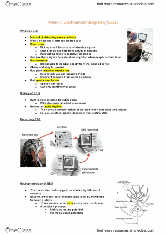
Recording EEG
Electrode cap
oArranged in circular grid
F: frontal; P: parietal; C: central; O: occipital; T: temporal
oAmplifier
oExperimental stimulation computer
oEEG recording computer
1
find more resources at oneclass.com
find more resources at oneclass.com

PSYC20006 Biological Psychology
Neurophysiology of EEG
EEG activity originates mostly from post-synaptic potentials
(voltages arising when NTs bind to receptors on membrane of post-synaptic cell)
oCauses ion channels to open/close
oLeads to graded changes in potential across membrane
oUnderstood as small dipole
Signals from single cells not strong enough to be recorded outside of
head
oIf many neutrons spatially align, summed potentials add to create recordable
signals
oPooled activity from groups of similarly oriented neutrons comes mostly from
large cortical pyramid cells
Functional unit = over 10,000 simultaneously activated neurons
Orientation of neurons determines sign of recorded potentials
oSome orientations lead to signals which can’t be recorded
Limitations
EEG biased to signals generated in superficial layers of cerebral cortex
on gyri directly bordering skull
oSignals in sulci harder to detect than gyri; maybe masked by gyri
Meninges, cerebrospinal fluid (CSF) & skull “smear” EEG signal
oDifficult to localise source
oInverse problem: mathematically, if sources known, scalp configuration
of signals can be reconstructed, but reverse untrue
A given configuration can have multiple dipole solutions
Analysing EEG data
The signal
oMeasured in relation to reference electrode
Reference should be neutral point, e.g. tip of nose, mastoids
oTypical amplitude of 10 -100 micro V (tiny)
Need to be amplified 1000-100,000x
oSignal then digitalised
Typical sample frequency: 256-1024Hz but up to >4000Hz
oBand-pass filtered to remove low (<0.5-1Hz) and high (>35-70Hz) frequency
because they cannot reflect brain activity
EEG is the sum of signals originating from many different neural units
Processing data
EEG signal very noisy, so to clean:
oRaw signal
oFiltering
oEpoching (divide into chunks time locked to interesting parts)
oBaseline correction
oArtefact rejection
Artefacts: everything that could change the signal / aren’t brain signals
2
find more resources at oneclass.com
find more resources at oneclass.com
Document Summary
Weeks 1 - 5: statistics & imaging methods. Place electrodes on scalp o o o o o o o. Electrodes pick up small fluctuations of electronic signals. Raw signals recorded are very noisy and don"t look like much. Can use signals to learn things about cognition when people perform tasks. Can do intra-cranial eeg, measuring directly at exposed cortex. Great temporal resolution (measure things quickly as they change) Disadvantage: spatial resolution not great (can"t zoom" in) Hans berger detected first eeg signal in 1924. Electrodes attached to scalp of his wife, reported results in 1929. When people close their eyes, an electrical signal varies with a characteristic frequency (8-13 hz) o. 1 at front of head, 1 at rear. F: frontal; p: parietal; c: central; o: occipital; t: temporal. Eeg activity originates mostly from post-synaptic potentials (voltages arising when nts bind to receptors on membrane of post-synaptic cell) o o o. Leads to graded changes in potential across membrane.