BIOL1003 Lecture Notes - Lecture 3: Haversian Canal, Hyaline Cartilage, Epiphyseal Plate

19 May 2018
School
Department
Course
Professor

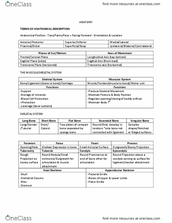
When body is standing erect, eyes looking forward, arms at sides of the body and palms and toes
directed forwards
-
Anatomical position
Body planes
Sagittal
Divides body into right and left parts.
Midsagittal
Divides body into equal halves
Transverse
Divides body into superior and inferior parts
Coronal
Divides the body into anterior and posterior
Anatomical terms
elongated body of long bone.
DIAPHYSIS -
proximal and distal expanded ends of long bone.
EPIPHYSIS -
where bones form joints.
ARTICULAR SURFACES -
smooth, nearly flat, articular surface.
FACET -
rounded articular projection.
CONDYLE -
joint between bones formed by fibrocartilage.
SYMPHYSIS -
large, distinct bony projection.
PROCESS -
slender, often pointed, bony projection.
SPINE -
shallow depression.
FOSSA -
opening in bone, a hole.
FORAMEN -
canal-like passageway.
MEATUS -
SINUS -
Anatomy and terms
Sunday, 26 March 2017
2:43 PM
3. The Skeletal System Page 1

cavity within a bone, filled with air.
SINUS -
3. The Skeletal System Page 2

Describe the anatomy and structure of bone
206 bones
•
Bone is living tissue
•
Bone = connective tissues
•
Bone
○
Cartilage - connective tissue which combines strength and flexibility, flexibility due to lack of mineral salts which are present in
bone
○
Skeleton consists of 2 types of connective tissue
•
Both consist of fibres and cells surrounded by a matrix, which is secreted by the cells
•
Rigid, dense connective tissues
•
Matrix is strengthened by the deposition of salts e.g. calcium carbonate and calcium phosphate
•
Gives rigid support and protection
•
Main tissue of the axial and appendicular skeletons of adults
•
Living tissue - never static
•
Constantly being eroded away/rebuilt by cells within the bony tissue
•
Anatomy of bone
Cell types
Production of bone matrix
•
Osteoblasts - build
○
Receive nutrients and eliminate wastes
•
Uses a system of canal containing blood vessels connected to the circulation
•
Substances diffuse in and out through the canaliculi
•
Osteocytes - maintain
○
Removal or resorption of bone
•
Osteoclasts - breakdown
○
Composition (in
extracellular
matrix)
25% organic collagen (tough, ropelike protein), give flexible strength to bone, elasticity and strength, secreted
by osteoblasts on surface of developing/growing bone
-
50% inorganic minerals e.g. calcium, phosphate, give bone compression/weight-bearing strength
-
25% water
-
4 shapes - long, short, flat, irregular
•
Long
Longer than they are wide
-
Most bones of the upper and lower limbs
-
E.g. femur
-
Short
Approximately wide as they are long
-
E.g. bones of the wrist and ankle
-
Flat
Thin flattened shape
-
E.g. certain skull bones, ribs, scapulae, sternum
-
Irregular bones
Vertebrae
-
Facial bones
-
Shapes do not fit into other 3 categories
-
Structure of bone
Long bone structure
Useful for illustrating parts of a typical bone
•
Diaphysis
Central shaft
Epiphysis
2 ends
-
Thin layer of articular cartilage covers ends of epiphyses where bone joins with
other bones
-
Epiphyseal plate/growth
plate
Composed of cartilage
-
Located between epiphysis and diaphysis
-
Only present if bone still growing
-
This is where bone grows in length
-
When bone growth stops, cartilage replaced by bone and become epiphyseal
line
-
Cavities
Filled with fatty yellow bone marrow
•
E.g. large medullary cavity in diaphysis
-
Smaller cavities in epiphyses of long bones and interior of other bones
-
Yellow marrow - consists mostly of adipose tissue
•
Red marrow - consists of blood-forming cells and only site of blood
formation in adults, gives rise to WBC
•
As person ages, red marrow mostly replaced by yellow marrow
•
Cavities filled with MARROW (soft tissue)
-
Periosteum
Consists of 2 layers
-
Contains blood vessels and nerves
-
Dense connective tissues
-
Covers most of the outer surface of bone
-
Endosteum
Lines surface of medullary cavity
-
Thinner connective tissue membrane
-
Articular cartilage
Thin layer of cartilage at ends of long bones that articulate with each other at
the joints
-
Types of bone
Compact (outer)
Mostly solid matrix and cells
•
Solid matrix - gives strength rigid
•
Haversian system - control canal,
canal iculi, lacunae
•
Forms most of the diaphysis of a long
•
3.1
Sunday, 26 March 2017
2:28 PM
3. The Skeletal System Page 3
Document Summary
When body is standing erect, eyes looking forward, arms at sides of the body and palms and toes directed forwards. Epiphysis - proximal and distal expanded ends of long bone. Symphysis - joint between bones formed by fibrocartilage. Sinus : the skeletal system page 1. Sinus - cavity within a bone, filled with air: the skeletal system page 2. Skeleton consists of 2 types of connective tissue. Cartilage - connective tissue which combines strength and flexibility, flexibility due to lack of mineral salts which are present in bone. Both consist of fibres and cells surrounded by a matrix, which is secreted by the cells. Matrix is strengthened by the deposition of salts e. g. calcium carbonate and calcium phosphate. Main tissue of the axial and appendicular skeletons of adults. Constantly being eroded away/rebuilt by cells within the bony tissue. Uses a system of canal containing blood vessels connected to the circulation. Substances diffuse in and out through the canaliculi.