CAM101 Lecture Notes - Lecture 30: Streptococcus Pyogenes, Hair Follicle, Skin Biopsy


Infection
Transmission
Epidemiology
Clinical Manifestation
Diagnosis
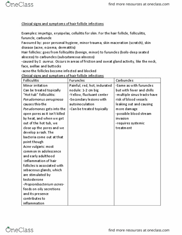
Folliculitis
S. aureus
Bacteria gaining entry through
abrasions
Single or multiple reddish lesions, can be
tender or painless, and can appear on and part
of hair-bearing skin
Made on clinical
presentation
Furuncles
• S. aureus
• Occasionally S.
pyogenes
Bacteria gaining entry through
abrasions
Deep infection of a hair follicle. Lesion appears
red at first, giving rise to a large (1 to 2cm in
diameter) pus-filled painful fluctuant nodule
Made on clinical
presentation
Carbuncles
• S. aureus
• Occasionally S.
pyogenes
Bacteria gaining entry through
abrasions
Multiple boils (or infections involving several
hair follicles) coalesce giving rise to multiple
abscesses
Made on clinical
presentation
Impetigo
• Crusted
• S. aureus
• S. pyogenes
Penetrates intact skin or
through previous trauma
Yellow crusted ulcerative erosions that are itchy
but generally not painful
Culture of skin
swab
Impetigo
• Bullous
S. aureus
Penetrates intact skin or
through previous trauma
Large blisters, initially filled with clear fluid that
later become cloudy and yellow, rupture easily
and brownish crusts form
Culture of skin
swab
Erysipelas
Group A S. pyogenes
Gain entry through break in
skin
Rapidly spreading bright red, raised, firm and
swollen plaque, with sharp demarcated borders
Made on clinical
presentation
Cellulitis
• S. pyogenes
• Occasionally S. aureus
N/A
Shows typical signs of inflammation; redness,
swelling, warmth, and pain
• Culturing
aspirates (pus)
of the lesion
• Skin biopsy
Pityriasis Versicolor
(aka Tinea Versicolor)
Malassezia furfur
Colonising the outer
(keratinised) layers of skin, hair
and nails
Small hypo/hyper-pigmented macules on any
part of the body, macules are irregular in
shape, raised and sometimes covered by fine
layer of scales
Microscopic
examination of
skin scrapings
Cutaneous Mycoses
(commonly named
ringworms or tinea)
-Tinea Capitis
Three fungi species
• Microsporum
• Trichophyton
• Epidermophyton
Numerous, mainly from
animals
Tinea Capitis (Scalp)
• Primarily a childhood disease, associated
with inflammation and scaling
Tinea Barbae (Beard)
• Microscopic
examination of
skin scrapings
find more resources at oneclass.com
find more resources at oneclass.com
Document Summary
Impetigo: bullous, aureus, occasionally s. pyogenes, aureus, occasionally s. pyogenes, aureus, pyogenes, aureus. Single or multiple reddish lesions, can be tender or painless, and can appear on and part of hair-bearing skin. Lesion appears red at first, giving rise to a large (1 to 2cm in diameter) pus-filled painful fluctuant nodule. Multiple boils (or infections involving several hair follicles) coalesce giving rise to multiple abscesses. Yellow crusted ulcerative erosions that are itchy but generally not painful. Large blisters, initially filled with clear fluid that later become cloudy and yellow, rupture easily and brownish crusts form. Rapidly spreading bright red, raised, firm and swollen plaque, with sharp demarcated borders. Shows typical signs of inflammation; redness, swelling, warmth, and pain. Colonising the outer (keratinised) layers of skin, hair and nails. Small hypo/hyper-pigmented macules on any part of the body, macules are irregular in shape, raised and sometimes covered by fine layer of scales: culturing aspirates (pus) of the lesion.