ANHB2214 Lecture Notes - Lecture 15: Portal Vein, Hepatic Artery Proper, Porta Hepatis

26 Mar 2020
School
Course
Professor
Document Summary
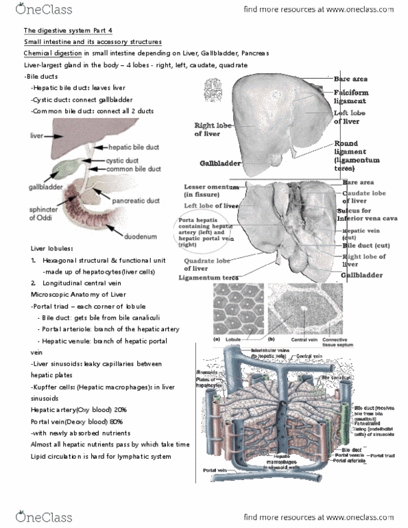
Liver: all venous drainage from the gastrointestinal tract passes through the liver, git, pancreas, gallbladder, spleen. Liver sinusoids: largest internal organ= 2% of body weight, upper right quadrant, below diaphragm, has major left and right lobes with, 2 smaller interior lobes covered by thin capsule and mesothelium of visceral. Ligament that contains obliterated umbilical vein: falciform ligament, peritoneal reflection off anterior abdominal wall with round ligament in its margin, ligamentum venosum: Ligamentous remnant of fetal ductus venosus, allowing fetal blood from placenta to bypass liver. It can become engorged: pain in right hypochondrium, runner"s stitch, enlarged liver easily palpable inferior to right costal margin. Thick fibrous connective tissue capsule covered with serous visceral peritoneum. Bile duct- of cuboidal epithelium, branches of bile conducting system. Includes stellate sinusoidal macrophages (kupffer cells) are a regular part of the hepatic sinusoid lining. They are members of the mononuclear phagocytic system and are derived from monocytes.