RIU 332 Lecture Notes - Lecture 14: Left Gastric Artery, Common Iliac Artery, Esophageal Hiatus

13 Mar 2020
School
Department
Course
Professor
Document Summary
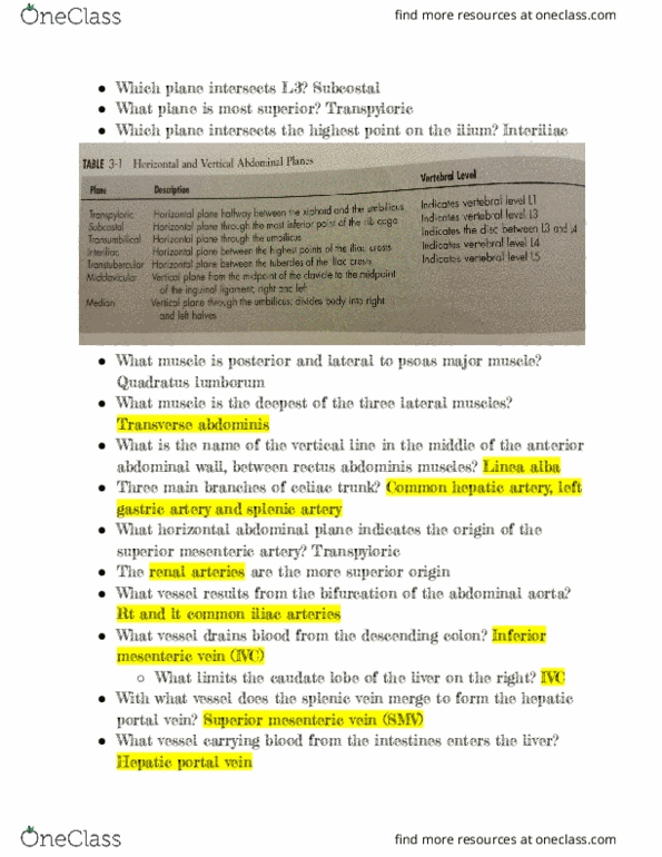
After its formation, what direction does the portal vein follow, to the right. Two branches of the celiac trunk are shown in this figure. Hepatic veins take blood out of the liver to the ivc. Two vessels carry blood into the liver. Hepatic artery has higher oxygen than portal vein. The aortic hiatus is located at the level of the tenth thoracic vertebra. The kidneys, the pancreas, and stomach are retroperitoneal. The renal arteries are paired and arise from the aorta at the level of the third lumbar vertebra. Imv, splenic vein, portal vein, liver sinusoids, central vein, and hepatic vein. On the visceral surface, the right lobe is separated from the rest of the liver by the ivc and the gallbladder. The flexure between the duodenum and jejunum is attached to the anterior abdominal wall by a peritoneal ligament. The left suprarenal gland is related to the hepatic flexure of the colon and to the left kidneys.