11:680:390 Lecture Notes - Lecture 2: Gram Staining, Antimicrobial Resistance, Gram-Negative Bacteria

13 Mar 2018
School
Department
Course
Professor

Document Summary
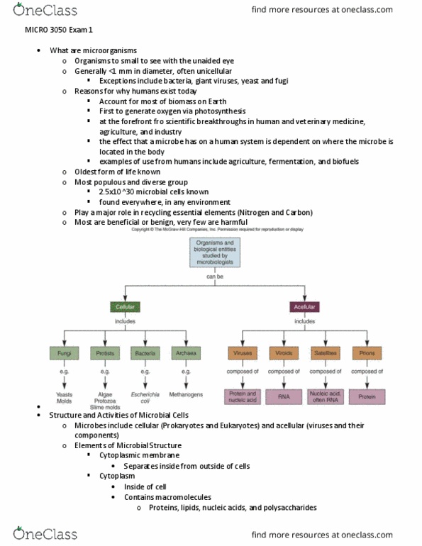
2: microbial cell structure and function: organelles that all living cells have, cell membranes, ribosomes, dna, properties of all cells, metabolism: cells take up nutrients, transform them, an expel wastes. Tools of microbiology: microscopy cheetah!!: bacteria/archaea: 0. 5 - 2 um, viruses: ~50-100 nm, light microscopy, magnification: the ability to make an object appear larger, resolution: the ability to distinguish two adjacent objects as separate and distinct. Limit of resolution for light microscope = 0. 2 um. Improving contrast in light microscopy (1) gram stain: coat the outside of the cell; can have positive or negative chronoform (a) thinner cell wall: pink (b) thicker cell wall: purple: bright-field microscopy, phase-contrast microscopy. Exploits differences in the refractive index of cell and surroundings (light refracts from organism) Allows for visualization of live samples: dark-field microscopy. Image appear light on a dark background. Excellent for observing motility: fluorescence microscopy. Cells appear to glow on black background due to filters: electron microscopy, tem.