KNES 364 Lecture Notes - Lecture 24: Qrs Complex, T Wave, Graph Paper

25 Mar 2020
School
Department
Course
Professor
Document Summary
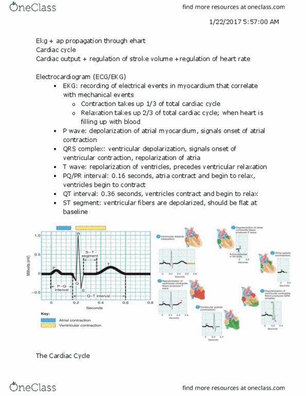
Getting a permanent recording as it goes through the cardiac cycle. Qrs complex= atria repolarizing/ ventricles depolarizing (ventricles are largest muscle mass of heart) T wave= ventricles repolarizing (even before the t wave) J point all through st segment and t wave= ventricles repolarizing. Printed on specific paper (graph paper) designed for ekg because it is more helpful. Smaller boxes= . 04 seconds beg end of box. Qrs does not look normal (double peak) t wave looks upside down. 3*. 04= . 12 taking more time because of blocked area (disorder l. bundle branch block) ** at max should take . 1 seconds to depolarize rs: Important to place in specific locations to be able to compare the 2 ekgs to one another. Lead- camera angle or a certain view of the heart. For this class putting on chest for safety rather than legs and arms since it is involving exercising. #1 looking from ra electrode to la electrode (straight across)