PSIO 532 Study Guide - Midterm Guide: Macrophage, Epithelium, Pressure Measurement


PSL – Respiratory Physiology
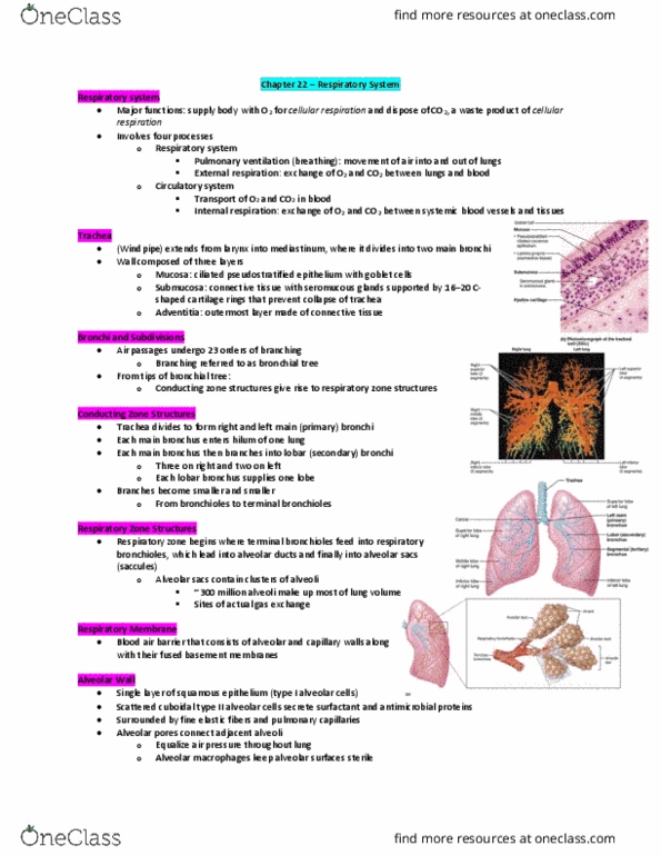
Types of Alveoli & Pressures
o Alveoli are surrounded by elastic fibers and an
extensive network of capillaries; They contain open
alveolar pores to equalize pressure and have alveolar
macrophages to phagocytize particulate matter that
may be drawn into the alveoli
o Elastic fibers → allows the alveoli to expand and
contract (allows us to maintain the integrity of the
lung); the elasticity of the fibers would no longer exist
if one was to smoke for a long period of time →
diminish the ability for your lungs to recoil
o Alveolar Macrophages: another protective
mechanism at the level of the alveoli → traps the
foreign particles, put them into the lymphatic system,
which helps us eliminate the foreign particle
o Alveoli has 2 types of cells: type-I & type-II
o The respiratory membrane consists of a single layer of squamous epithelium,
type-I cells, surrounded by a basal lamina.
o Interspersed among the type-I cells are cuboidal type-II cells that secrete
surfactant.
o Type-I: primary involved in gas exchange
o Type-II: produces surfactant (surfactant reduces the surface tension that exists
at the level of the alveoli)
o Alveolar Pores: these pores help the alveoli to interact with one another; so if
one alveolus expands, it promotes the expansion of the other alveoli that
surrounds that particular alveolus (interconnection between the alveoli) → helps
the lung to work in concert during expansion
find more resources at oneclass.com
find more resources at oneclass.com
Document Summary
Pressure differences across the lung are dependent on the coupling of the lung (green) and chest wall (black line border); the blue is the intrapleural space. When the lungs are uncoupled from the chest wall they recoil to minimal volume (lungs alone) less than residual volume; this is a resting position of the lungs. Residual volume (rv) the volume left in the lungs after blowing out air as much as possible. Since our lung is coupled to the chest wall, it would never collapse down to a lower level; it would only go down to the level of residual volume. At resting frc, the pressure of intrapleural space is -5 cmh2o (average value between top and bottom of the lung) this value is standardize to atmospheric pressure. Since atmospheric pressure is 760, the absolute pressure value in the intrapleural space would be 755 cmh20. Uncoupling of the lung from the chest wall is known as a pneumothorax.