BIOL 3500 Chapter Notes - Chapter 11: Serous Gland, Pancreatic Duct, Pancreatic Islets

22 Oct 2016
School
Department
Course
Professor

Document Summary
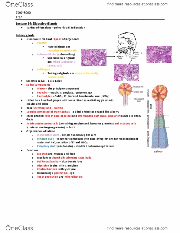
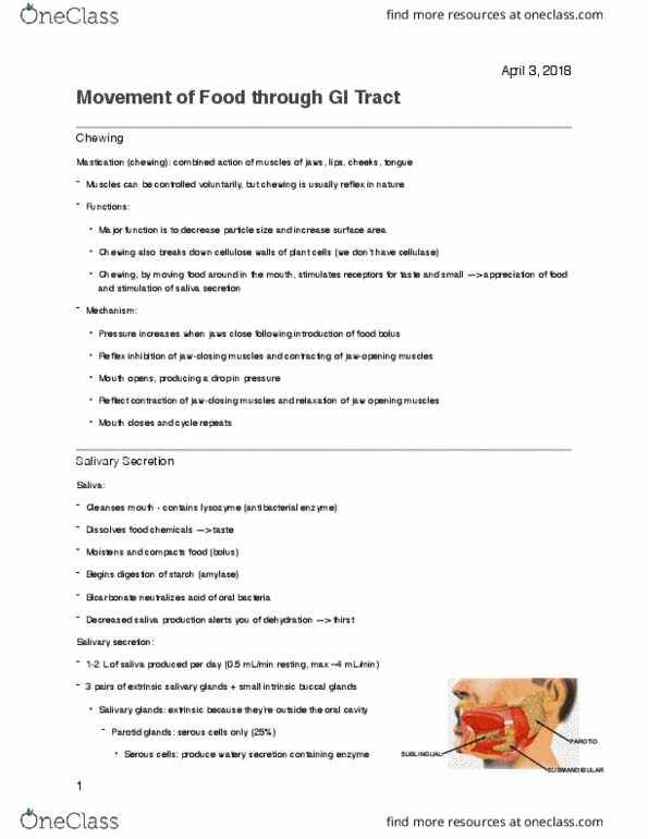
Antibacterial agents connective tissue capsule lobules, separated by septa originating from capsule www. webmd. com courses. stu. qmul. ac. uk/smd/kb/microanatomy/sdentists/ob2/8. htm. White in h&e --> mucus does not stain. Duct portions intercalated ducts smallest branches attached to acini single cuboidal ep striated ducts single cuboidal to low columnar ep. Myoepithelial cells (basket cells) around acinar and duct cells cytoplasmic processes. Myoepithelial cells in this section from salivary gland can be shown by an immunoperoxidase technique that demonstrates desmin, which is the muscle-speci c intermediate lament and stains brown. Parotid glands largest of the salivary glands thick capsule and septa: mostly serous secretory cells, amylase, antimicrobial proteins. Submandibular (submaxillary) glands both mucous and serous some mucous portions capped by serous demilunes amylase anti bacterial proteins. Some capped by serous demilunes. produces mucus. Secrete digestive enzymes into duct system: endocrine cells: islets of langerhans. Exocrine pancreas: branched tubuloacinar serous gland, duct system intercalated ducts (low cub ep) lobular ducts (columnar ep)