MCB 2610 Chapter Notes - Chapter 1B: Chromophore, Formaldehyde, Microscope Slide

8 Sep 2016
School
Department
Course
Professor
Document Summary
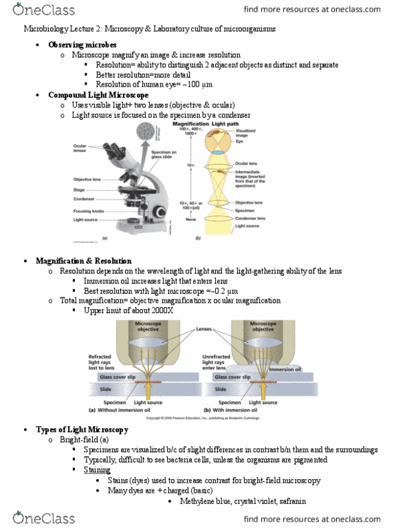
Avery welch mcb 2610 microbiology week 2 & 3 chapter 1 appendix b microscopy & specimen prep: specimens usually stained with fluorochromes (fluorescent dyes) Fluorescein isothiocyanate (fitc) often attached to antibodies that bind specific cellular components or to dna probes. Fluorochrome-labeled probes, such as antibodies ,or fluorochromes tag specific cell constituents for id of unknown pathogens. Can be used to antibody staining in strains like streptococcus pyogenes: other uses for fluorescence microscopy. Photosynthetic organisms naturally fluoresce when exited with specific wavelengths. Can also be used to localize specific proteins within cells. One method is to fuse the gene of the protein of interest to the jellyfish (aequorea) green fluorescent protein (gfp) Can be fused the mbl cytoskeletal protein of bacillus subtilis. Confocal microscopy: confocal scanning laser microscopy (clsm) creates sharp, composite 3d image of specimens using laser beam, aperture to eliminate stray light, and computer interface, specimen is usually fluorescently stained, numerous applications including study of biofilms.