ALHT106 Lecture Notes - Lecture 9: Natural Killer Cell, Memory T Cell, Blood Vessel


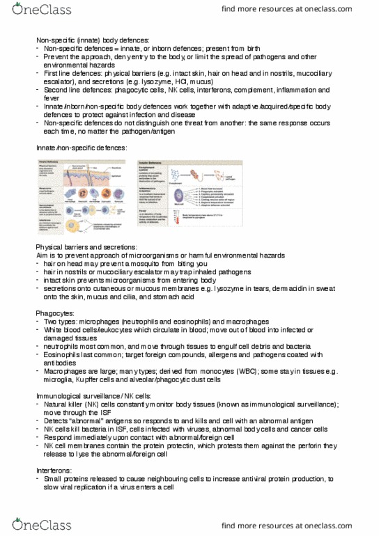
The lymphatic system:
-the lymphatic system includes the cells, tissues and organs which defect the body against
pathogens (disease-causing microorganisms)
-Primary cells of the lymphatic system are lymphocytes:
-B cells
-T cells
-Natural killer (NK) cells
-NK cells part of non-specific body defences (2nd line of defence)
-B and T cells part of specific body defences (3rd line of defence)
-1st of defence is preventing the pathogen from entering the body
Immunity:
Immunity= ability to resist infection and disease
-Two forms of immunity: innate (nonspecific) and adaptive (specific)
-Lymphocytes (B and T cells) within lymphoid organs and tissues defend the body against
invading pathogens and abnormal or cancerous body cells
-Macrophages/phagocytic cells present also in lymph tissues and organs to engulf and destroy
foreign/abnormal/cancerous cells
Lymphatic system structures:
1. Lymph: fluid similar to plasma but with much lower protein concentration
2. Lymphatic vessels/lymphatics: network of vessels starting in tissues > ducts that empty into the
subclavian veins
3. Lymphoid tissues and organs scattered throughout the body
4. Cells: B and T cells, dendritic cells and phagocytes
Function of the lymphatic system:
1. Produce, maintain and distribute lymphocytes that defend against infection and environmental
hazards e.g. toxins; lymph tissues and organs
2. Carry lipids/chylomicrons absorbed from the GIT to the blood
3. Maintain normal fluid balance and ISF composition in peripheral tissues (returns cleaned fluid
to blood)
Lymphatic vessels 1:
-Smallest vessels are lymphatic capillaries
-starts as pockets
-Large diameters
-thin walls
-Walls of endothelium supported by an incomplete basement membrane and regions of overlap
-One way valves allow fluids, solutes as large as proteins, viruses, bacteria and cell debris to
enter but not to return to ISGG
-Lymphatic capillaries lie alongside blood capillaries
-From capillaries, lymph flows into larger vessels; large lymph vessels contain many valves to
prevent back flow of fluid to peripheral tissues; valves cause bulge in vessel
-Superficial lymphatics in subcutaneous layer; drains arteriolar tissue of mucous and serous
membranes
-Deep lymphatics drain skeletal muscles, walls of visceral organs, and organs of the trunk, neck
and limbs
-Deep and superficial lymphatics join to form large lymphatic trunks- thoracic or lymphatic ducts
Lymphocyte:
Three types of lymphocytes circulate in blood:
1. T cells (Thymus dependent cells)
2. B cells (Bone marrow derived cells)
3. Natural Killer (NK) cells
find more resources at oneclass.com
find more resources at oneclass.com

-Most are T cells: Cytotoxic T (Tc) cells, Helper T (Th) cells, Memory T cells and Suppressor T
(Ts) cells
-T cells responsible for cellular.cell-mediated immunity
-B cells responsible for antibody mediated/humoral immunity
-NK cells part of innate/inborn immunity; immunological surveillance
-Most have long lifespans: 4-20+ years; produced in bone marrow and lymphoid tissues and
organs
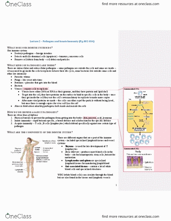
Lymph tissues and organs:
-CT dominated by lymphocytes; often contained in lymph nodules
-Lymph nodules have densely packed lymphocytes in an area of areolar CT
-Lymph nodules in tonsils (guard entrance to respiratory and digestive tracts); other collections in
respiratory and digestive tracts; also in urinary and reproductive tracts
-Lymph organs are surrounded by a protective, fibrous, CT capsule which separates them from
surrounding tissues
-Lymph organs, lymph nodes, the thymus and the spleen
MALT and tonsils:
-Mucosa-Associated Lymphoid Tissues = MALT are the clusters of lymph nodules that protect the
epithelia of the digestive, urinary, respiratory and reproductive tracts
-Examples of MALT are Peyer’s patches in intestinal walls, the appendix and the tonsils
-Appendix: blind pouch originating near junction of small and large intestine; contains lymph
nodules in the walls
-Tonsils in the pharynx walls; most people have five (5) tonsils (L & R palatine, one large
pharyngeal, and L & R lingual)
-If infected > tonsillitis with inflammation including swelling
Lymph nodes:
-Small organs (1-25mm); mainly located in axillary, inguinal and neck regions
-Lymph nodes shaped like kidney beans with a hilum/ indentation
-Afferent lymphatics take fluid into nodes and efferent lymphatics lead away from the nodes
-Function to filter/purify the lymph from toxins, viruses, foreign and abnormal cells, cancerous
cells and cell debris
-As lymph returns to the blood it has had 99% of all antigens removed
-Dendritic cells start an immune response, and macrophages engulf pathogens and act as
antigen-presenting cells (APCs) to B and T cells
Thymus:
-Located in the mediastinum, posterior to the sternum
-Thymus is largest relative to body size in babies and infants (maximum around 2 years old);
increases in size until puberty then decreases and becomes more fibrous
-T cells in cortex actively divide and cells move to medulla to mature under the influence of
thymosins
-T cells enter the bloodstream and circulate around the body
Spleen:
-Contains the largest collection of lymphoid tissue in the body
-Perform the same functions for blood as lymph nodes do for lymph
-Functions:
-Removal of abnormal blood cells and cellular components from blood by phagocytes
-Storing iron recycled from erythrocytes
-Initiating immune responses by B and T cells in response to circulating antigens
-Tears easily even though surrounded by a capsule
Summary and questions:
-The lymphatic system functions in body defences; other functions also. What are they?
find more resources at oneclass.com
find more resources at oneclass.com

-Lymphatic vessels collect ISF and lymph nodes filter antigens out of fluid; 99% of antigens
removed when returned to blood. How do antigens get removed?
-Collections of lymph tissues guard the digestive, respiratory, urinary and reproductive tract
epithelia. What are some examples?
-What are the major functions of the lymph nodes, thymus and spleen?
Roles of inflammation:
-Inflammation: protective response intended to:
4. Eliminate initial cause of cell injury
5. Remove damaged tissue
6. Prepare area for growth of new tissue
-Inflammation occurs before tissue repair, and replacement of damaged tissue or creation of scar
tissue, at the site of an injury or infection
Causes of inflammation:
-Immune response to infectious microbes
-Trauma
-Surgery
-Caustic chemicals
-Extremes of heat and cold
-Ischemic damage to body tissues
Inflammation and disease:
-Inflammation can also be present with disease in the body
-e.g. inflammation plays a well established role in bronchial asthma and autoimmune diseases
such as Rheumatoid arthritis
-Increasing evidence suggests inflammation also plays a role in atherosclerosis, diabetes mellitus
and Alzheimer’s disease
-When inflammation is present, the disease name has “itis” at the end e.g. dermatitis = skin
inflammation; arthritis = joint inflammation
Two inflammation patterns:
1. Acute inflammation
-Relatively short duration; nonspecific; early response to injury
-Aims are to remove the injurious agent, and to limit tissue damage
2. Chronic inflammation
-Longer duration, lasting for weeks to years
-Recurrent or progressive acute inflammatory process, or a low-grade smouldering response that
does not evoke an acute response
Chronic inflammation:
-Typically caused by low-grade, persistent, infections or irritants, which are unable to penetrate
deeply or spread rapidly. Also present in autoimmune diseases due to ongoing tissue attack
-E.g. foreign bodies such as Talc, Silica, Asbestos, Surgical material
-Low virulence Viruses, Bacteria, Fungi
-Large parasites with moderate to low virulence such as TB or Syphilis
find more resources at oneclass.com
find more resources at oneclass.com
Document Summary
The lymphatic system includes the cells, tissues and organs which defect the body against pathogens (disease-causing microorganisms) Primary cells of the lymphatic system are lymphocytes: Nk cells part of non-speci c body defences (2nd line of defence) B and t cells part of speci c body defences (3rd line of defence) 1st of defence is preventing the pathogen from entering the body. Two forms of immunity: innate (nonspeci c) and adaptive (speci c) Lymphocytes (b and t cells) within lymphoid organs and tissues defend the body against invading pathogens and abnormal or cancerous body cells. Macrophages/phagocytic cells present also in lymph tissues and organs to engulf and destroy foreign/abnormal/cancerous cells. Walls of endothelium supported by an incomplete basement membrane and regions of overlap. One way valves allow uids, solutes as large as proteins, viruses, bacteria and cell debris to enter but not to return to isgg.