BIOL125 Lecture Notes - Lecture 5: Endomysium, Ultimate Tensile Strength, Epimysium


- Muscle cells (like neurons) have excitability/irritability due to a polarised membrane
ability to contract/shorten and relax/lengthen movement in body
- Note: muscle cell = muscle fibre; contractile proteins known as (myo)filaments
- Muscle cells tend to have prefix “sarco” ie.
oSarcolemma = plasma/cell membrane
oSarcoplasm = cytoplasm
oSarcoplasmic reticulum = endoplasmic reticulum – like organelle
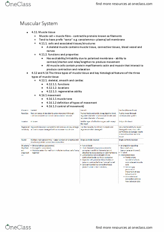
- Three types: skeletal, cardiac and smooth muscle cells (and tissues)
- Each have different locations in the body; all create movement by shortening upon
contraction and lengthening upon relaxation
- Cardiac and smooth cells have a single nucleus in contrast to skeletal cells
- Smooth muscle twists upon contraction while cardiac and skeletal muscles directly
shorten
- All 3 types of muscle cells contain protein myofilaments: actin and myosin (with
varying arrangement) that interact to produce contraction and relaxation
- Skeletal muscle cells are multinuclei
Properties of Skeletal Muscle
- Both voluntary and autonomic
(involuntary) control
- Contraction under control of somatic
motor neurons
- Somatic = body
- Multinucleate ie. Formed from
multiple myoblast cells fusing
together
- Multiple nuclei large numbers of
enzymes and structural proteins
needed for contraction
- Many mitochondria; lots of ATP
needed for contraction
mitochondria on and around the
contractile filaments
- Striated arrangement: myofibrils arranged in sarcomeres
- Q. Provide examples of involuntary/autonomic contraction of skeletal muscle
protective type of responses eg: eye twitch; autonomic control of regulating of
breathing at night
- Striations thin filaments =
- Intercolated disc allows rapid transfer of ions through cells
- Cardiac cells have intercolated discs – bottom picture on slide 7
- Q. What capacity for regeneration do they have? Depends on ability to perform
mitosis
oCardiac muscle cells have limited capacity for regeneration. Cells become
hypertrophic to generate extra force
oSkeletal muscle cells cannot undertake mitosis – ability to repair and
regenerate because of myosatellite cells?
oSmooth muscle cells – when we have enough cells, they will stop dividing. But
this can be reactivated to replace damaged smooth muscle cells
find more resources at oneclass.com
find more resources at oneclass.com

Origin and insertion of a Skeletal Muscle
- Muscles attach to bones via tendons in two places: a fixed point and a moveable
point
- Origin point of tendon attachment to the less moveable bone; ie. Bone does not
change position during muscle contraction; typically proximal to insertion
- Insertion point of tendon attachment on the more moveable bone, ie. Moves bone
during contraction; located across the other side of the joint from the origin
- When a muscle contracts, the insertion point moves towards the point of origin
- Muscles can only pull bones, not push them, so they work in antagonistic pairs or
sets
- Have to have crossing over a joint to allow flexion and extension
Skeletal muscle attachment to bone
- Skeletal muscles attach to bone via:
1. Tendons
2. Flat/broad sheets (aponeuroses)
- These attachments are primarily collagen fibres (as in ligaments) which extend into
the bone providing a firm attachment (collagen fibres have high tensile strength)
- Tendon: extension of collagen fibres of the epimysium, perimysium and endomysium
which together form a bundle
- Aponeurosis: collagen fibres forming a flat/broad sheet
- Attachment points on bones are rough to allow strong attachment
- Muscle is made up of bundles (fascicles) of muscle fibres surrounded by perimysium
for protection
- Each muscle cell is surrounded by endomysium which isn’t as tough as perimysium
to allow blood vessels to get through to muscle cell
- How the fascicles are arranged determines the type of movement
- Myofilaments (actin and myosin) arranged into sarcomeres
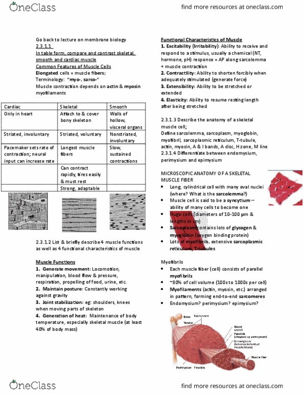
Internal Organisation of a skeletal muscle fibre/cell
- Myofibrils: run entire length of cell; organised in repeating end-to-end
structural/contractile units (sarcomeres); attached to sarcolemma at both ends of
muscle fibre; external part of
sarcolemma attached to
collagen fibres of tendon
- When myofibril
shortens/contracts, entire
muscle fibre shortens so pulls
on tendon movement
- Myofibrils surrounded by
mitochondria and glycogen
(reserve for ATP formation)
- Myofibrils = 2 contractile
myofilaments/proteins;
actin/thin filament and
myosin/thick filament; plus other proteins (titin, troponin and tropomyosin)
- Myofibrils regulate how much contraction is needed
- If actin side is covered, no contraction can occur
find more resources at oneclass.com
find more resources at oneclass.com
Document Summary
Muscle cells (like neurons) have excitability/irritability due to a polarised membrane. Ability to contract/shorten and relax/lengthen movement in body. Note: muscle cell = muscle fibre; contractile proteins known as (myo)filaments. Muscle cells tend to have prefix sarco ie. sarcolemma = plasma/cell membrane: sarcoplasm = cytoplasm, sarcoplasmic reticulum = endoplasmic reticulum like organelle. Three types: skeletal, cardiac and smooth muscle cells (and tissues) Each have different locations in the body; all create movement by shortening upon contraction and lengthening upon relaxation. Cardiac and smooth cells have a single nucleus in contrast to skeletal cells. Smooth muscle twists upon contraction while cardiac and skeletal muscles directly shorten. All 3 types of muscle cells contain protein myofilaments: actin and myosin (with varying arrangement) that interact to produce contraction and relaxation. Multinucleate ie. formed from multiple myoblast cells fusing together. Multiple nuclei large numbers of enzymes and structural proteins needed for contraction.