BIOL126 Lecture Notes - Lecture 1: Sclera, Itch, Cyanosis

8 Jun 2018
School
Department
Course
Professor

Integumentary System
• Functions
o Protection of underlying tissues from impact damage, abrasion, fluid loss and
chemicals
o Maintenance of normal body temperature via insulation or evaporative cooling
o Synthesis of Vitamin D3 by UV light -> converted to calcitriol by liver for absorption
of calcium and phosphorous from the GIT
o Detection of the external environment via thermoreceptors (heat and cold),
mechanoreceptors (fine touch, pressure, vibration receptors) and nociceptors (pain)
o Production of keratin to protect against abrasion and provide water resistance
(multiple layers of dead, waterproofing cells)
o Production of melanin (pigment) by melanocytes which protects underlying tissue
from UV radiation
o Storage of lipids in adipocytes in dermis and adipose layer of hypodermis
o Excretion of salts and water by sweat glands; and excretion of oils for lubrication
(and release of small amounts of organic wastes) by sebaceous glands
• Name the components of the integumentary system and describe their main functions
o Two major components
• Cutaneous membrane
▪ 1.5-2m2 in area, accounting for about 16% of body weight
▪ First line of defence to protect body against external environment, and
informs brain about the environment
▪ Intact skin:
• Reduces water loss
• Prevents pathogen entry
• Protects against impact, chemicals and UV light
▪ Has two portions (epidermis and dermis)
• Accessory structures
▪ Hairs
▪ Follicles
▪ Sweat glands
▪ Sebaceous glands
▪ Nails
▪ Arrector pili muscles
find more resources at oneclass.com
find more resources at oneclass.com

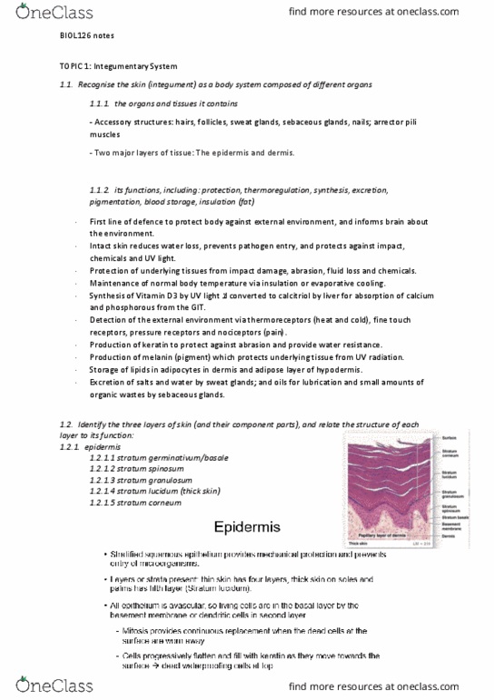
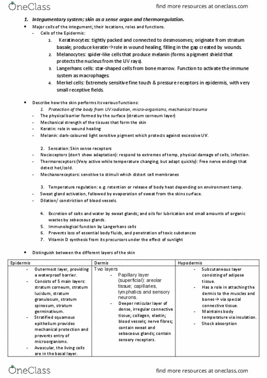
o Identify the three layers of skin (and their component parts), and relate the
structure of each layer to its function
• Epidermis
▪ Stratified squamous epithelium
• Dermis
▪ Areolar/loose connective tissue
▪ Dense, irregular connective tissue
• Hypodermis (subcutaneous adipose layer)
▪ Separates integument from deep fascia and other organs
• Connective tissue fibres of hypodermis interweave with those of the dermis
▪ Holds tissue layers together
• Epidermal ridges and dermal papilla increase SA for interconnections between
epidermal and dermal layers
o Summarise the structure and function of the epidermis, dermis and hypodermis
• Epidermis
▪ Stratified squamous epithelium provides mechanical protection and
prevents entry of microorganisms
▪ Layers of strata present
• Thin skin has four layers, thick skin on soles, palms and fingertips
has fifth layer (stratum lucidum) - outer layer much thicker
▪ All epithelium is avascular so living cells are in the basal layer by the
basement membrane or dendritic cells in second layer
• Mitosis provides continuous replacement when the dead cells at
the surface are worn away
• Cells die, progressively flatten and fill with keratin as they move
towards the surface -> 15-30 layers of dead waterproofing cells at
top
▪ Cell types
• Basal cells
• The germinative cells which undergo mitosis to replace
continual loss of upper layer cells
• Keratinocytes
• Most common
• Filled with keratin protein
• Flatten and die as they progress outwards
• Outermost layer is 15-30 layers of dead keratinocytes tightly
connected by desmosomes
• Melanocytes
• Pigment-producing cells in basal layer (skin and hair colour)
• Merkel cells
• Tactile cells scattered in basal layer (of skin without hair)
find more resources at oneclass.com
find more resources at oneclass.com

• Dermis
▪ Two layers
• Superficial papillary layer of areolar tissue; capillaries, lymphatics
and sensory neurons
• Deeper reticular layer of dense, irregular connective tissue;
collagen, elastin; blood vessels; nerve fibres; contain sweat and
sebaceous glands; contain sensory receptors
▪ Collagen
• Great tensile strength
• Some give but resists stretching, pulling and twisting
• Alignment of collagen fibres along tension lines allows skin to resist
force applied during normal movement
▪ Elastin
• Gives stretch and recoil ability to skin
▪ Skin turgor
• Water present which gives resilience and flexibility to skin
▪ Role of dermal circulation
• Oxygenated blood looks bright red since Hb bound to oxygen ->
oxyhaemoglobin
• As oxygen is released and more Hb is formed, blood gets a darker
red-blue colour -> red in arterial blood and blue in venous blood
as seen through the skin
• Vasodilation of blood vessels in dermis
• Increases blood flow through capillaries -> pink coloured
skin and greater conduction of heat through the skin i.e.
greater heat loss
• Vasoconstriction
• Heat conservation and pale skin
• Skin easily observed and can aid diagnosis of body conditions
find more resources at oneclass.com
find more resources at oneclass.com
Document Summary
First line of defence to protect body against external environment, and informs brain about the environment. Intact skin: reduces water loss, prevents pathogen entry, protects against impact, chemicals and uv light, has two portions (epidermis and dermis, accessory structures. Sebaceous glands: hairs, nails, arrector pili muscles. Identify the three layers of skin (and their component parts), and relate the structure of each layer to its function: epidermis. Stratified squamous epithelium: dermis, areolar/loose connective tissue, dense, irregular connective tissue, hypodermis (subcutaneous adipose layer) Stratified squamous epithelium provides mechanical protection and prevents entry of microorganisms. Functions: protect from uv damage, help cushion light impact, reduce insect and pathogen entry e. g. on head, in nostrils, eyelashes, eyebrows stop sweat from entering eyes. Follicle: surrounded by connective tissue then sensory neuron (root hair plexus) Sebaceous glands: oil glands, discharge oily, lipid secretion (sebum) into hair follicles and onto skin.