PHTY100 Lecture Notes - Lecture 3: Internal Obturator Muscle, Greater Trochanter, Anterior Superior Iliac Spine


3. Introductory myology and muscles of the hip region
• 3.1 Identify and describe the major components of a skeletal muscle organ – belly,
attachments, connective tissue
o Belly - contractile cells, where the work happens
o Attachment to the skeleton
• Must cross a joint
• Proximal attachment called the 'origin'
• Distal attachment called the 'insertion'
o Connective tissue
o Muscle tissue
• Muscle cells called muscle fibres, which do the work
• Capillary network - requires a rich blood supply for the rapid delivery of
nutrients and removal of wastes
• Fibrous connective tissue
▪ Allows the work of the cell to be transferred to the muscle organ for
movement
▪ Consists of the epimysium, perimysium and endomysium
o Muscle structure
• Myofibres - muscle cells
▪ Cells fuse together
▪ Multi-nucleated
▪ Striations
• Myofilaments - strands of protein within the muscle cells
• Actin and myosin filaments slide over one another to bring about muscle
shortening
▪ Filaments do not shorten
▪ Receptor sites on the actin called troponin
▪ Calcium binds onto the troponin, changing its shape and opens the
binding site for myosin to latch onto the actin
▪ ATP releases energy to bind and drag the thin filament over the thick
find more resources at oneclass.com
find more resources at oneclass.com

find more resources at oneclass.com
find more resources at oneclass.com

• 3.2 Describe the different types of attachments of skeletal muscle organs – tendinous,
fleshy, raphe
o Fleshy
• Muscle fibres attach directly to the bone, via a very small amount of
connective tissue
o Tendon
• Muscle fibres attach to a cord of connective tissue, which then attaches to the
bone
o Raphe
• Muscle fibres attach to a sheet of connective tissue, which attaches to
another muscle
• Gives muscles something to attach onto when there isn't a bone
o Aponeurosis
• Sheet of connective tissue from muscle to bone
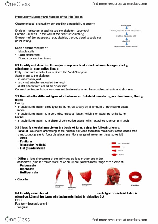
• 3.3 Classify skeletal muscle on the basis of form, using the following terms:
o Parallel - fibres run in a straight line between attachments
• Maximum shortening of the muscle belly and therefore movement at the
associated joint
• Not great for force development
• More range of movement, less powerful
• Fusiform
▪ Narrowed fibres at each end
▪ Biceps brachii
o Oblique - fibres run on an angle between attachments
• Less shortening of the belly and so less movement at the associated joint
• More power, less range of movement
• Look like feathers
• Unipennate
▪ Fibres on one side of the tendon
• Bipennate
▪ Fibres coming from 2 directions to the axis
• Multipennate
▪ Sections of bipennate
find more resources at oneclass.com
find more resources at oneclass.com
Document Summary
Fibrous connective tissue: allows the work of the cell to be transferred to the muscle organ for movement, consists of the epimysium, perimysium and endomysium, muscle structure, myofibres - muscle cells, cells fuse together, multi-nucleated. Striations: myofilaments - strands of protein within the muscle cells, actin and myosin filaments slide over one another to bring about muscle shortening. Fusiform: narrowed fibres at each end, biceps brachii, oblique - fibres run on an angle between attachments. Less shortening of the belly and so less movement at the associated joint: more power, less range of movement, unipennate. Fibres on one side of the tendon: bipennate. Fibres coming from 2 directions to the axis: multipennate. Sections of bipennate: circular - fibres run around an opening, 3. 4 identify examples of each type of skeletal listed in objective 3. 3 and the types of attachments listed in objective 3. 2, types of attachments.