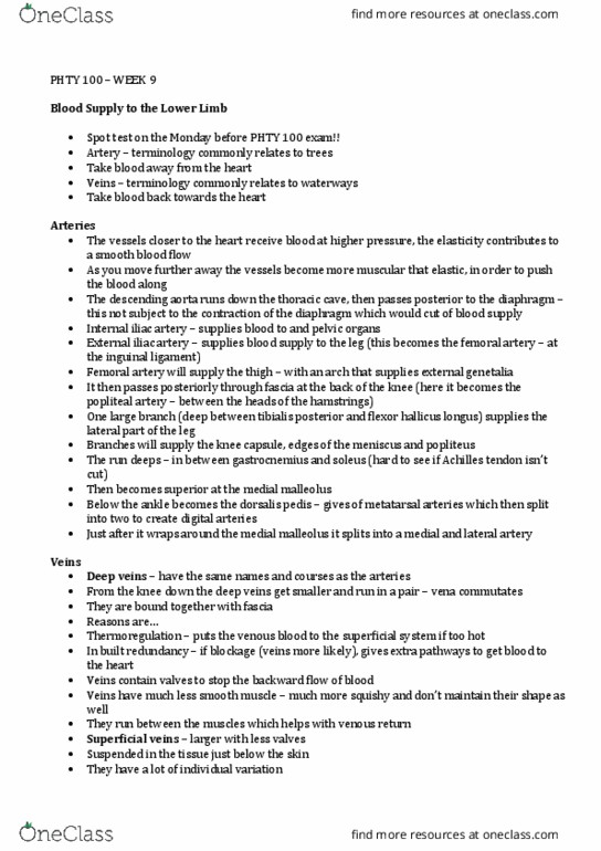
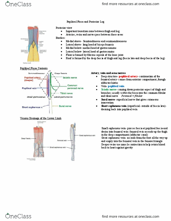
PHTY100 Lecture Notes - Lecture 10: Posterior Tibial Artery, Anterior Tibial Artery, Internal Iliac Artery

Document Summary
Branches of abdominal aorta: left and right common iliac arteries, divide into internal iliac artery and external iliac artery, becomes femoral artery once it passes under the inguinal ligament. Arteries of lower limb: femoral artery, profunda femoris, popliteal artery, anterior tibial artery. Continues into dorsal aspect of foot: peroneal artery. Supplies lateral compartment: posterior tibial artery. Two sets: deep veins, run with and take the names of the arteries, same pattern as arteries, vena commitantes, contain valves, superficial veins: In the skin: valves (not as many as deep, communicate freely with deep veins, best identified in surface anatomy. Significance of two sets: two ways to return blood to heart, some in built redundancy, temperature control. Segmented innervation of lower limb and nerve lesions. Damage to peripheral nerves: most vulnerable sites: Between skin and bony projection- crushing injury. Myotomes: group of muscles supplied by one spinal nerve, often described in terms of movements that the muscles bring about.